Creative Biolabs offers blocking assays (also known as antibody competition assays) to help clients study cancer antigens and identify immunodominant epitopes. These assays are useful for classifying antibodies and controlling allergic disorders.
Learn More →BAFF Assay Portfolio Service
Background Molecules What We Can Offer Why Choose Us FAQs Customer Review Related Services Contact Us
Introduction
B cell-activating factor (BAFF), also known as B lymphocyte stimulator (BLyS) and tumor necrosis factor (TNF) ligand superfamily member 13B (TNFSF13B), is a cytokine belonging to the TNF ligand family that activates B cells. BAFF is first synthesized as type II transmembrane proteins, mainly in myeloid and stromal cells, and further processed by furin convertase cleavage into trimeric soluble cytokines BAFF 3-mer. Twenty soluble BAFF 3-mers can be further oligomerized into a virus-like particle, called BAFF 60-mer. BAFF expression is increased in the presence of type I interferon (IFNs) including IFN-γ, interleukin (IL)-10 and granulocyte colony-stimulating factor, as well as by Toll-like receptor 3 (TLR3), TLR4 or TLR9 stimulation.
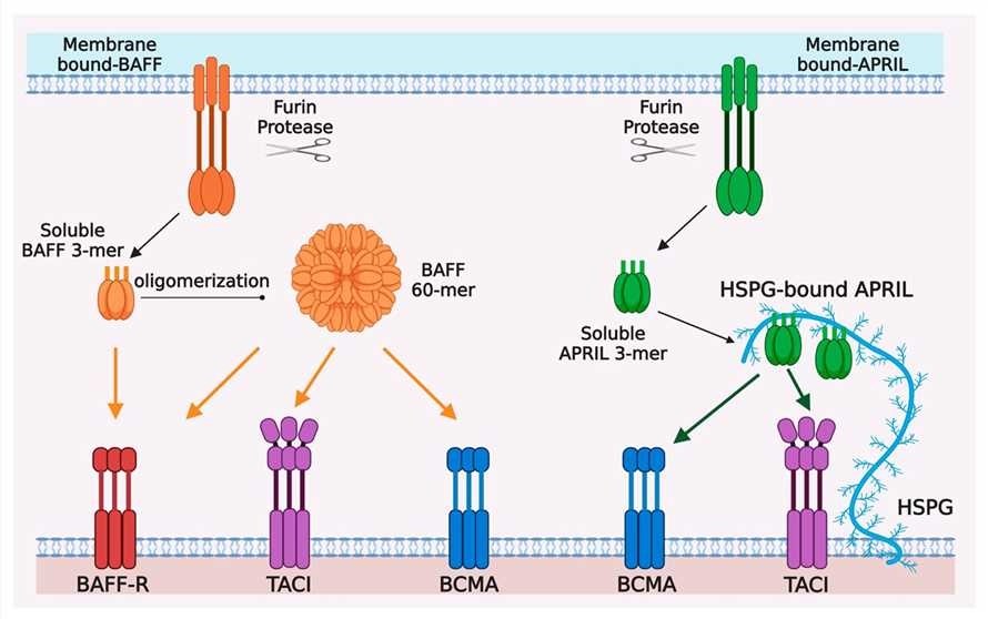 Fig.1 Ligand-receptor interactions in the BAFF/APRIL system.1, 3
BAFF System Molecules
The so-called "BAFF system molecules", which consist of two ligands, BAFF and a proliferation-inducing ligand (APRIL), and three receptors, are critical for B cell homeostasis. BAFF and APRIL are type II transmembrane proteins and they share some receptor specificity. BAFF binds to 3 receptors that are primarily expressed on B cells: BAFF receptor (BAFF-R), B cell maturation antigen (BCMA), and transmembrane activator and CAML interactor (TACI). Both BAFF and APRIL bind two receptors BCMA and TACI. APRIL does not bind BAFF-R but binds heparan sulfate proteoglycan (HSPG) in both the extracellular matrix and on the surface of cells like plasma cells, triggering APRIL multimerization. The expression of all three receptors is restricted mainly to B lymphocyte lineage cells. BAFF and APRIL are cytokines that share biological functions, they promote B cell survival and maturation and are expressed as membrane-bound or soluble proteins.
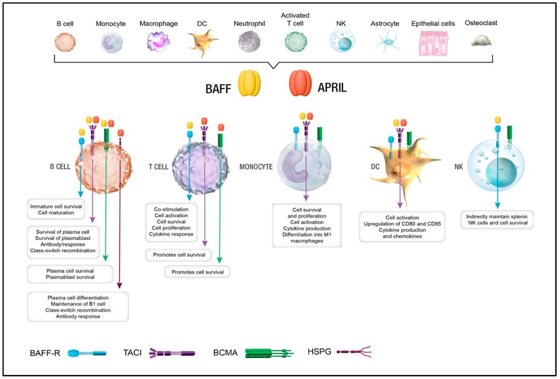 Fig.2 The role of BAFF and APRIL in regulating immune homeostasis.2, 3
What We Can Offer?
BAFF is considered an important player in many pathophysiological conditions, including inflammation, autoimmune disorders, and primary immunodeficiencies. Increased expression and serum levels of BAFF were demonstrated in many types of hematological and solid tumors, making BAFF a possible new biomarker in malignancies. Creative Biolabs provides a full set of BAFF assay portfolio services for research:
- NF-κB activation assay
- In vitro B-cell stimulation assay
- ELISA assay for BAFF
- Cell proliferation assay
- Cell apoptotic assay
- Cell migration assay and wound healing assay
- Immunofluorescence and immunohistochemistry
- Flow cytometry: cell surface antigens and apoptosis
- Western blotting
Contact us today to discuss how our services can support your project.
Why Choose Us?
With deep expertise in immunobiology and drug development, Creative Biolabs delivers cutting-edge BAFF assay services to accelerate your research. The BAFF system—including receptors BAFF-R, BCMA, and TACI—plays a vital role in B-cell survival and is linked to autoimmune diseases and cancers like multiple myeloma. Studies highlight TACI as a promising therapeutic target, especially for overcoming antigen escape in CAR-T therapies.
Our tailored solutions combine advanced technology with flexible customization, ensuring precise support for your project. Whether exploring drug discovery or immunotherapy, we provide the tools and insights to advance your work. Partner with us to unlock new possibilities in targeting the BAFF pathway for innovative treatments.
Frequently Asked Questions
Q1: How do you ensure the reproducibility of your assays?
A1: We use standardized operating procedures (SOPs) and rigorous quality control on all reagents and assays to ensure high reproducibility. Our final report includes a detailed methodology for transparency and replicability.
Q2: Can your assays handle novel or custom-designed compounds?
A2: Our platform is highly flexible for custom assay development. We adapt protocols to accommodate a wide range of compounds, including small molecules, bispecific antibodies, and CAR-T cells.
Q3: I am concerned about the confidentiality of my proprietary compounds and data. How do you handle that?
A3: We adhere to the highest standards of data security and confidentiality. All projects are conducted under a strict confidentiality agreement, and your materials and data are handled with the utmost care by our dedicated, expert team.
Customer Review
-
Actionable Insights
The flow cytometry analysis from Creative Biolabs provided crucial, actionable insights into TACI and BCMA expression on our patient-derived cells, validating our multi-target therapeutic strategy. - S. H. K***h
-
Comprehensive Data
The comprehensive and high-quality data from Creative Biolabs' ELISA service, with its exceptional reproducibility, was perfect for our regulatory submission and superior to off-the-shelf kits. - A. L. P***d
Related Services
To further support your research, we offer a range of complementary services that can be integrated with your BAFF assay project.
B Cell-based Cross Blocking Assay
Binding Affinity and Specificity Evaluation Service
Creative Biolabs offers antibody-directed enzyme prodrug therapy (ADEPT) in vitro services, utilizing advanced biophysical techniques and high-throughput screening to assess antibody-enzyme binding.
Learn More →How to Contact Us
Creative Biolabs has been dedicated to providing one-stop tumor marker assay services (e.g., BAFF assay portfolio service). We are experienced in the specialized development of high-complexity assay solutions to support complete custom solutions for biochemical and cell-based assays. This facilitates pharma and biotech preclinical research in all therapeutic areas and the development of companion diagnostics and clinical biomarkers, particularly in oncology. If you are interested in our services, please feel free to contact us.
References
- Xu, Shengli, and Kong-Peng Lam. "Transmembrane activator and CAML interactor (TACI): another potential target for immunotherapy of multiple myeloma?" Cancers 12.4 (2020): 1045. DOI: https://doi.org/10.3390/cancers12041045
- Ullah, Md Ashik, and Fabienne Mackay. "The BAFF-APRIL system in cancer." Cancers 15.6 (2023): 1791. DOI: https://doi.org/10.3390/cancers15061791
- Distributed under Open Access license CC BY 4.0, without modification.
 Download our brochure
Loading case studies...
Online Inquiry