Flow Cytometry-based ATM and H2A.X Assay Service
DNA Damage Signals: ATM and H2A.X
ATM (Ataxia Telangiectasia Mutated) and H2A.X (a variant of the histone H2A) are crucial players in the DNA damage response pathway. ATM is a serine/threonine protein kinase that plays a pivotal role in the detection and repair of DNA double-strand breaks (DSBs). They are two critical biomarkers directly involved in the apoptotic pathways. Upon sensing DNA damage, ATM rapidly phosphorylates histone variant H2A.X at the serine 139 residue, forming γ-H2A.X, a molecular signal that facilitates the recruitment of DNA repair proteins to the site of damage. Together, they serve as essential indicators for monitoring cellular responses to DNA damage and the subsequent initiation of apoptosis. Given their significance, precise detection and quantification of ATM and H2A.X are pivotal for both research and therapeutic applications. At Creative Biolabs, we offer a comprehensive flow cytometry-based ATM and H2A.X assay designed to accurately assess and quantify these key molecular indicators.
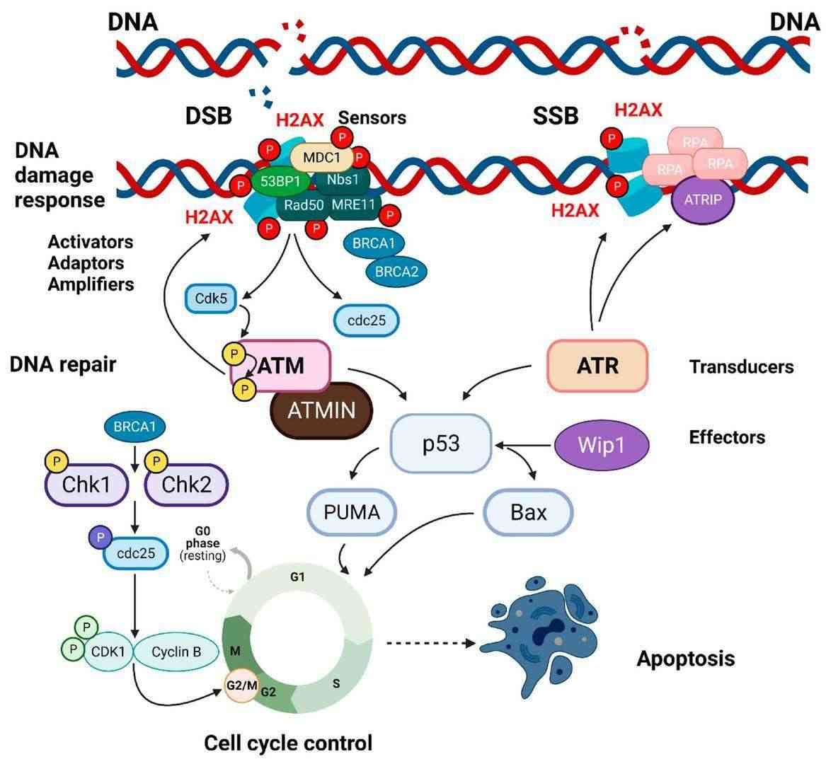 Fig.1 Simplified pathways of DNA damage response.1
Flow Cytometry-based ATM and H2A.X Assay for Assessing Late Apoptosis
Flow cytometry stands out as a powerful and versatile tool for single-cell analysis, enabling precise measurement of protein expression levels and cell cycle status within heterogeneous cell populations. At Creative Biolabs, we leverage advanced flow cytometry techniques to offer precise detection and quantitative analysis of ATM and H2A.X activation and phosphorylation states in cells. Our assay is designed to provide detailed insights into the stages of apoptosis and DDR, allowing researchers to track how cells respond to various treatments or environmental conditions.
The process begins with sample preparation, where cells are harvested, fixed, and permeabilized to allow intracellular staining. We use highly specific antibodies conjugated with fluorescent dyes that bind to ATM and γ-H2A.X, ensuring that the detection is both accurate and reliable. Once the cells are prepared and appropriately stained, they are analyzed using a flow cytometer. The flow cytometer utilizes laser excitation and advanced optics to detect the fluorescence signals emitted by the antibody-dye complexes. Our data acquisition software then translates these signals into quantitative data, offering a detailed profile of ATM and γ-H2A.X expression levels among the cell populations. This approach not only allows us to distinguish between apoptotic and non-apoptotic cells but also to dissect distinct stages of apoptosis and DNA damage responses with unparalleled precision.
Applications
- Drug Development: Screening potential drug candidates that induce apoptosis through DNA damage.
- Toxicology: Studying the genotoxic effects of environmental toxins and other hazardous substances.
- Cancer Research: Evaluation of chemotherapeutic efficacy by monitoring DNA damage and apoptosis in cancer cells.
- Molecular Biology: Investigating the molecular mechanisms underlying DNA repair and cell cycle control.
Explore more about our services and discover how Creative Biolabs accelerates your research by contacting us today.
Reference
- Merighi, Adalberto, et al. "The phosphorylated form of the histone H2AX (γH2AX) in the brain from embryonic life to old age." Molecules 26.23 (2021): 7198. Distributed under Open Access license CC BY 4.0, without modification.
 Download our brochure