B Cell-based Immunohistochemistry Assay
Principal of Immunohistochemistry Assay
The immunohistochemistry (IHC) technique is a powerful approach to determining locations of certain protein distribution characters in tissues based on specific antibodies' recognition of the corresponding antigen proteins on formalin-fixed paraffin-embedded tissues. It is easy to detect the primary antibody by an antibody binding to its Fc regions. Commonly, the second antibody conjugates with fluorochrome or enzyme as a reporter that facilitates visualization by imaging techniques, such as fluorescence microscope, light microscope, fluorescence electron microscope, and chemiluminescence reactions. Based on this advantage of immunostaining, Creative Biolabs offers antibody-mediated immunohistochemistry assays for cancer antigen studies.
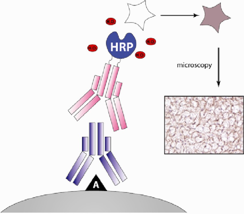 Fig. 1 Schematic representation of immunohistochemistry.1
IHC-based Cancer Epitope Analysis Service at Creative Biolabs
The immunohistochemistry technique can analyze specific proteins on intact tissues and cells, providing microscopic images based on immunostaining and chromogenic/fluorescent reaction. Creative Biolabs provides the whole process of services on immunohistochemistry, including sample preparation, antibody production, immunostaining, and data interpretation.
✔ Two Types of Staining
-
Both direct and indirect immunostaining are available.
-
Your type of staining is chosen based on the abundance of the antigen and accessibility.
We provide the best sample collection, preservation, and fixation approaches to maintain tissue structural integrity and state. It can be fixed in formalin and then embedded in paraffin wax or frozen to make histologic sections for antibody-based staining.
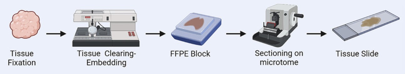 Fig.2 Tissue slide preparation.3,4
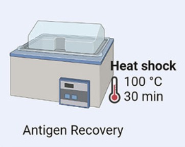
✔ Antigen Recovery
Antigen retrieval techniques are applied to uncover antigen epitopes buried due to sample fixation and processing, including enzyme-dependent proteolysis and heat-induced disassociation of linked proteins.
✔ Antibody
Source of Antibodies
-
Your antibodies of interest;
-
Customized antibody production;
-
Select from our high-quality antibody library validated with a clear target range and standard staining protocols.
Various Antibody Types
We provide mouse, humanized, chimeric monoclonal antibodies, and Fab fragments, that can be produced and used for immunohistochemistry and immunocytochemistry to capture your target of interest.
Secondary antibodies are optimized for high specificity without binding to endogenous antibodies.
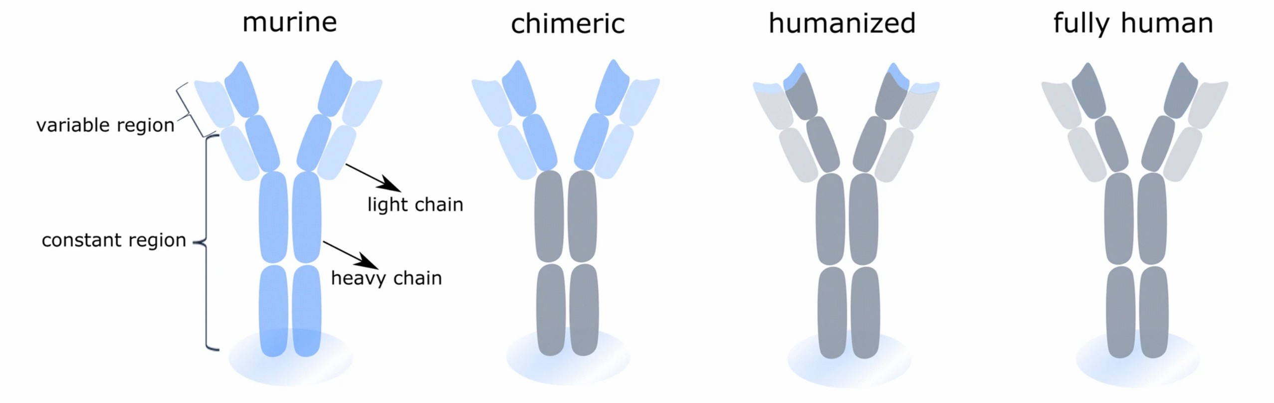 Fig.3 Types of antibodies for analysis.2,4
✔ Detection Approaches
A Fluorescence microscope is used to detect fluorescent staining. Chromogenic staining, including polymer-based detection and biotin-based detection, and the former is recommended for increased sensitivity, is detected by light microscopy.
We also provide counterstain assay for clients to visualize cellular anatomy and differentiate specific staining.
✔ Data Report
Staining Results Can Be Reported in Types
-
Qualitative
-
Semi-quantitative
-
Quantitative
The Digital Detection System
-
Quantitative detection
-
Increased sensitivity and accuracy
-
Capable to detect rare events
Highlights
-
Customized assay design.
-
Data analyzed by pathologists combined computing algorithms.
-
Fast delivery and cost-effective.
If you are interested in our immunostaining assays, please contact us for further details.
References
-
Jensen, L. D., et al. "Animal models of angiogenesis and lymphangiogenesis." Biomedical Science, Engineering and Technology. IntechOpen, 2012. Distributed under Open Access license CC BY 3.0, without modification.
-
Van Hoecke, Lien, and Kenny Roose. "How mRNA therapeutics are entering the monoclonal antibody field." Journal of Translational Medicine 17.1 (2019): 54.
-
Maiques, Oscar, and Victoria Sanz-Moreno. "Multiplex chromogenic immunohistochemistry to stain and analyze paraffin tissue sections from the mouse or human." STAR protocols 3.4 (2022): 101879.
-
Distributed under Open Access license CC BY 4.0, without modification.
For Research Use Only | Not For Clinical Use
 Download our brochure