Circuit-induced Cell Death Quantification Service
Quantifying cellular response to therapeutics is essential to understanding their mechanisms of action and assessing therapeutic efficacy. Directly measuring both cell growth and death can provide valuable information for interpreting the response of cells to the treatments. Creative Biolabs has developed several efficient approaches to more precisely quantify circuit-induced cell death, to further clarify the nature of cancer cell response and interactions.
Methods for Cell Death Detection
It is very important to analyze cell death processes that determine the cell death rate and sensitivity. There are several conventional methods to examine the cell death rate, such as cell survival, clonogenic, and membrane permeability assays. The colorimetric substrate is used to assess metabolic activity such as mitochondrial succinate dehydrogenase activity which depicts cell survival. That is why this method is considered a cell survival assay. However, clonogenic assays are typically used to stain colonies of cells that proliferate from cells that are resistant to or recovered from a cell death challenge and retain proliferation potential. Therefore, cells arrested in the G1 or G2 phase cannot be examined through clonogenic assays although the cells do not undergo cell death. Therapeutics or targeted genes-induced cell death may affect metabolism or proliferation without inducing death, which sometimes leads to misinterpretation using metabolic readouts without actually reflecting cell death induction. Alternatively, membrane permeability assays are usually used which are considered more reliable as they measure the end stage of the cell death process itself.
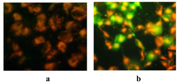 Fig.1 JC-1 dye accumulates in the mitochondria of healthy cells as aggregates (red-orange fluorescing). (Kari, et al., 2022)
Circuit-induced Cell Death Quantification Service at Creative Biolabs
Our scientists are proficient at multiple Genetic Methods to engineering cancer cells for self-destruction purposes, including Retrovirus, Lentivirus, Adenovirus, Adeno-associated Virus, and CRISPR-based Methods. After this step, Ex Vivo Immune Reactivity, Toxicity, and Circuit Performance Assessment are needed to assess the effect of self-destruction cancer cells. Quantification of circuit-induced cell death is one of the methods for circuit performance assessment.
Creative Biolabs has developed several effective methods for measuring cell death-related parameters. Those methods rely on various technologies and can be distinguished by their specificity, sensitivity, limitations, precision, and throughput.
At Creative Biolabs, a plethora of protocols for studying cell death are now available, including:
Membrane permeability/damage detection methods
- Annexin V binding assay
- Lactate dehydrogenase assay
- Electrochemical methods
Mitochondrial damage/alteration detection methods
- MTT and XTT assay
- Mitochondrial membrane potential detection
- Mitochondrial activity of streptolysin O permeabilized cells assay
- Cytochrome c release detection
Caspase activity detection methods
- ELISA
- Fluorometric and colorimetric assays
- Immunohistochemical methods
- Laser and mass spectroscopic methods
p53 activity detection methods
- Functional analysis of separated alleles in yeast (FASAY)
- p53 protein analysis methods
DNA -based detection methods
- APO single-stranded DNA (ssDNA) assay
- Terminal deoxynucleotidyl transferase (TdT)-mediated d-UTP nick end labeling (TUNEL) assay
- In-situ end labeling technique (ISEL)
- Gel electrophoresis-based methods
- DNA-specific fluorochrome-based methods
With an experienced team of in-house multiplex experts in circuit-induced cell death quantification, Creative Biolabs provides our clients with highly customizable solutions. For more detailed information, please feel free to contact us or directly send us an inquiry.
Reference
- Kari, S.; et al. Programmed cell death detection methods: a systematic review and a categorical comparison. Apoptosis. 2022, 27(7-8): 482-508.
 Download our brochure