EGF-induced HaCaT Cell Migration Assay Service
Unlock the Secrets of Skin Cell Movement
Cell migration is a fundamental process in skin health, affecting wound healing, tissue repair, and various dermatological conditions. To explore these mechanisms, Creative Biolabs introduces the epidermal growth factor (EGF)-induced HaCaT cell migration assay. This assay focuses on the role of EGF in stimulating keratinocyte migration, offering crucial insights for research into skin regeneration and therapeutic development. By precisely analyzing how EGF influences HaCaT cells, our service provides essential data for advancing skin-related research.
Technical Methods
Our EGF-induced HaCaT cell migration assay employs a streamlined approach for assessing cell migration:
| Assay Type | Cell Type | Model | Detection Method | Measured Response |
|---|---|---|---|---|
| Functional | HaCaT | Agonist | High-content Imaging | Fluorescence |
a) Cell Seeding: HaCaT cells are plated into a 96-well plate with a central cell-free area.
b) Treatment: Test substances, including EGF, are added to each well. The cells are cultured under controlled conditions for 44 hours.
c) Imaging and Analysis: Following incubation, cell migration into the central zone is analyzed using confocal imaging. The extent of migration is quantified by measuring the area of closure in the images.
This method offers a precise assessment of how EGF and other substances affect the movement of keratinocytes.
Advantages
Our EGF-induced HaCaT Cell Migration Assay offers several significant benefits:
- High precision: Advanced confocal imaging ensures accurate and detailed measurement of cell migration, providing reliable data for your research.
- Efficient and scalable: Suitable for high-throughput screening, the assay supports the evaluation of multiple test conditions in parallel, enhancing research efficiency.
- Relevance to skin research: Focused on EGF-induced migration, the assay delivers valuable insights into keratinocyte behavior, essential for skin health and therapeutic studies.
- Flexible customization: The assay can be adapted to test various compounds, conditions, or concentrations, allowing for tailored experiments.
Publication Sharing
This study investigates the effects of radiofrequency electromagnetic fields on HaCaT cell migration and cytokine expression in an in vitro wound healing model. The research demonstrates that exposure to electromagnetic fields significantly influences HaCaT cell migration, which is crucial for wound repair processes. The findings reveal that these fields can modulate cell movement, potentially enhancing or impeding the wound-healing process. Additionally, the study assesses changes in cytokine expression associated with cell migration, providing insights into the underlying mechanisms of how electromagnetic fields affect keratinocyte behavior and skin regeneration.
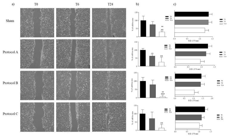 Fig.1 Wound healing assay.1
Applications
The EGF-induced HaCaT cell migration assay is ideal for:
- Wound healing studies: Assessing how EGF and other compounds affect skin cell migration, which is crucial for developing treatments for wound healing.
- Dermatological drug evaluation: Testing the efficacy of new drugs or compounds in influencing skin cell movement and proliferation.
- Basic research: Investigating the fundamental mechanisms of cell migration and the effects of various stimuli on keratinocyte dynamics.
Frequently Asked Questions (FAQs)
Q1: What specific cell line is used in the assay?
A: The assay utilizes HaCaT cells, a human keratinocyte cell line that is widely used for skin research.
Q2: What is the procedure for the assay?
A: HaCaT cells are seeded into a 96-well plate with a central cell-free zone. After adding the test substances and incubating for 44 hours, cell migration is assessed through confocal imaging.
Q3: What are the benefits of using EGF in this assay?
A: EGF is used as a positive control to stimulate cell migration, allowing us to compare the effects of test compounds against a known migratory response.
Q4: Can the assay be customized for different experimental conditions?
A: Yes, the assay can be tailored to include various conditions, compounds, or concentrations, providing flexibility for your specific research needs.
Q5: What type of data will be provided?
A: You will receive detailed quantitative data on cell migration, including high-resolution images and measurements of the migration area, to support your analysis.
For precise and insightful analysis of keratinocyte migration, EGF-induced HaCaT cell migration assay at Creative Biolabs is your go-to solution. Our expert team is ready to provide you with the high-quality data needed to advance your research in skin biology and therapeutic development. Contact us today to learn how our EGF-induced HaCaT cell migration assay can enhance your understanding of keratinocyte movement and propel your research forward.
Reference
- Costantini, Erica, et al. "Evaluation of cell migration and cytokines expression changes under the radiofrequency electromagnetic field on wound healing in vitro model." International Journal of Molecular Sciences 23.4 (2022): 2205. Distributed under Open Access license CC BY 4.0, without modification.
 Download our brochure