We offer specialized flow cytometry assays focused on characterizing B-cell populations, crucial for advancing epitope-based drugs, immuno-oncology, and autoimmune disease research.
Learn More →Immune Cell Surface Marker Identification Service
Creative Biolabs provides a definitive solution for researchers seeking to decipher the intricate language of the immune system. Our service delivers precise and comprehensive data on the identity and functional state of immune cells, empowering you to make data-driven decisions in your research. You can expect high-quality, multidimensional datasets that accurately profile cellular populations, identify key subpopulations, and reveal critical changes in response to various stimuli or treatments. Our outputs are tailored to support applications from target validation to biomarker discovery.
Introduction What We Can Offer Workflow Why Creative Biolabs Customer Reviews FAQs Related Services Contact Us
Immune Cell Surface Marker
Cell surface markers refer to membrane proteins embedded in the lipid bilayer structure of the cell membrane, including membrane antigens, membrane receptors, and other molecules. Cell surface markers are not only the material basis for mutual recognition between immune cells but also between cells or between cells and the matrix. Including receptors, MHC molecules, coordinated stimulation molecules, etc. The CD molecule is one of the most commonly used cell surface markers, which refers to the application of monoclonal antibody identification methods to identify the same differentiated antigen, including the specific morphology, specific function, and developmental stage of the cell.
Discover how our services can advance your research objectives - Request a consultation.
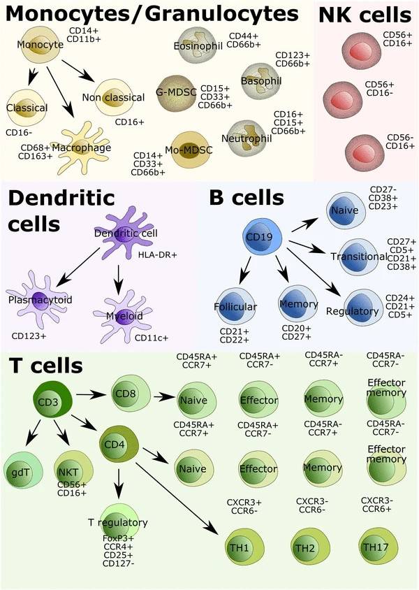 Fig.1 Some immune cells and their surface markers.1
What We Can Offer
-
Type of Our Methods
Creative Biolabs offers the following methods to help you identify the immune cell phenotype you need.
| Our Methods | Description & Features |
|---|---|
| Immunocytochemistry | The method is usually done with enzymes as antibody markers, using cellular enzyme immunohistochemistry. This method can be observed with a common microscope, and the stained cells are positive for the corresponding CD antigen. |
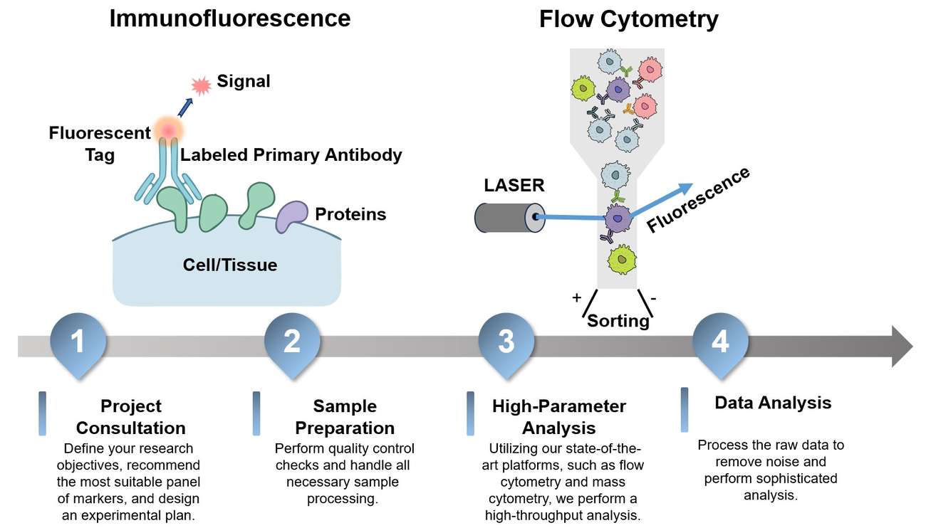
| Immunofluorescence | In this method, the isolated peripheral blood mononuclear cells are treated with the corresponding fluorescein-labeled CD monoclonal antibody, or with the corresponding CD monoclonal antibody, and the results are observed with a fluorescence microscope after preparation. |
| Flow cytometry | Flow cytometry is used for the detection of immune cells, and the results are objective, accurate, and reproducible. |
| Mass spectrometry | This method can achieve the analysis of ten protein biomarkers at the single-cell level and is widely used in the study of immune heterogeneity, tumor heterogeneity, etc. |
-
Type of Immune Cells
Immune cells perform specific functions in the immune system. Immune cell markers can be used to identify and distinguish between different types of immune cells. Below, we have listed common immune cells and their surface markers.
| Cell Type | Surface Marker |
|---|---|
| B cells | CD1c, CD10/Neprilysin, CD19, CD21, CD22, CD27, CD40, CD95, IgM, IgD, BCMA |
| Dendritic cells | CD11c, CLIP-170, Fascin, MADDAM, NLDC-145, MHCII (HLA-DR), CD33, CD123, CD16, CD128b |
| Granulocytes |
|
| Helper T Cells | BTLA, CD3, CD4, CD8, CXCL13, IFN-gamma, IL-4, IL-2, IL-5, IL-9, CCL17, CCL22, IL-21, IL-22 |
| Innate Lymphoid Cells | CD3, CD4, CD45, CD49a, CD69, IFN-gamma, TNF-alpha, ICOS, IL-4, IL-5, IL-9, GM-CSF, NKG2A |
| Macrophages | CD68, CD64, CSF-1R, F4-80 (mice), CD11b |
| Monocytes | CD14, CD16, CD115, Ly-6C |
| Myeloid-derived Suppressor Cells | CD11b/Integrin alpha M, CD14, CD15, CD11c |
| Regulatory T Cell | CD3, CD4, CD5, CD14, CD19, Galectin-1, IL-10, IL-35, GITR, TGF-beta |
Immune Cell Surface Marker Identification Service at Creative Biolabs
Highlights
Comprehensive Insights
We leverage proprietary technology that goes beyond conventional methods, allowing for the simultaneous detection of a far greater number of markers. This provides a high-resolution map of your cellular landscape for unmatched deep cellular insights.
Professional Expertise
Our dedicated team of specialists translates complex data into clear, actionable intelligence. We perform sophisticated analysis to remove noise from raw data and provide a precise understanding of your cellular populations and dynamics.

Precise and Reliable Results
Our platforms are designed for exceptional sensitivity and specificity. Through a rigorous process, we ensure accurate and reliable results, giving you the confidence needed to drive your research forward and make critical decisions.
Customized Solutions
At Creative Biolabs, our team works collaboratively with you to understand your unique research goals. We develop customized experimental plans and analysis strategies tailored to your specific project, ensuring the most relevant outcomes.
Discover how a partnership with us can streamline your research - Get a quote today.
Customer Reviews
-
Actionable Insights
The data from Creative Biolabs' service has greatly facilitated our research, providing us with the precise biomarker data we needed to stratify patients and understand mechanisms of resistance. - L***a T. -
Unmatched Detail
The high-parameter analysis provided by Creative Biolabs has allowed us to see cell populations we couldn't with our in-house flow cytometry, giving us an unprecedented level of detail in our cellular profiling. - A***d M.
FAQs
Do you offer services for specific cell types, such as T regulatory cells or myeloid-derived suppressor cells?
Yes, our service is highly customizable. We can design panels and analysis strategies to focus on a wide range of specific immune cell subsets and their activation states, as well as complex and rare cell populations. Please reach out to our team to discuss your specific cell type of interest.
How do you ensure the accuracy of your results?
We use advanced technology with exceptional sensitivity and specificity, along with rigorous quality control measures throughout the entire workflow, from sample preparation to data analysis. Our team of specialists meticulously processes and analyzes the data to ensure every cell is accurately profiled, providing you with reliable and reproducible results.
Related Services
B Cell-based Flow Cytometry Assay
B Cell-based Mass Spectrometry Assay
Our service utilizes mass spectrometry to determine the mass and amino acid sequence of epitope peptides, providing detailed insights into antibody-antigen binding for epitope mapping and characterization.
Learn More →How to Contact Us
Creative Biolabs has always provided expertise and timely communication throughout the service. If you are interested in our immune cell surface marker identification service, please feel free to contact us. We also offer more Bioassay services. If you are interested, please click to check it out!
Reference
- Lim, Su Yin, and Helen Rizos. "Immune cell profiling in the age of immune checkpoint inhibitors: implications for biomarker discovery and understanding of resistance mechanisms." Mammalian genome : official journal of the International Mammalian Genome Society vol. 29,11-12 (2018): 866-878. Distributed under an Open Access license CC BY 4.0, without modification. https://doi.org/10.1007/s00335-018-9757-4
 Download our brochure
Loading case studies...
Online Inquiry