T Cell based ³H-thymidine Assay Service
Background What We Can Offer Workflow Why Choose Us FAQs Customer Review Related Services Contact Us
Screening cancer epitopes, particularly neoepitope antigens, has become increasingly important in drug discovery and immunotherapy in recent years. Creative Biolabs provides various T and B cell-based epitope analysis assays, including T cell-based 3H-thymidine assay, to analyze cancer pathogenic epitopes.
Cancer Epitope and T Cell-based 3H-thymidine Assay
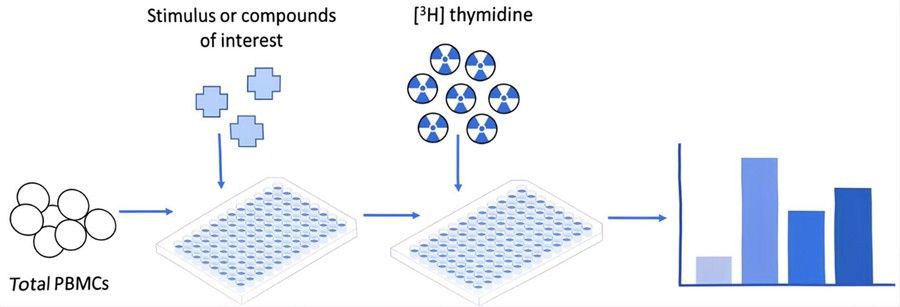
Cancer antigen epitopes are specific molecules that target T and B cell receptors and induce complex immune responses. Cancer epitopes are typically derived from somatic mutations that cause tumorigenesis and may progress to various diseases and cancer. T cell proliferation analysis is a reliable and reproducible method to determine the presence and frequency of epitope-specific T cells. The use of a 3H-thymidine incorporation assay to investigate T cell proliferative response is a validated tool to screen and test cancer pathogenic epitopes.
 Fig.1 Schematic representation of 3H-thymidine assay.1
A Comparative View of The Evolution of T Cell Proliferation Assays
While the 3H-thymidine assay has long been the gold standard, the field has evolved with the introduction of non-radioactive methods. The most notable alternative is the flow cytometry-based proliferation assay, which uses fluorescent dyes like carboxyfluorescein succinimidyl ester (CFSE) to track cell divisions.
Here is a brief comparison of the two methodologies:
|
3H-Thymidine Assay
|
Flow Cytometry Assay
|
-
Simplicity and High-Throughput
The 96-well format is easy to set up for large-scale screening.
-
Quantitative Data
Provides a highly reliable, singular measure of proliferation (CPM).
-
Cost-Effective
Often, it is more economical for high-throughput screens than flow cytometry.
|
-
Non-Radioactive
Eliminates the need for handling and disposal of radioactive materials.
-
Multi-Parameter Analysis
Allows for simultaneous measurement of proliferation alongside other parameters, such as cell surface markers, providing a richer data set.
-
Live-Cell Analysis
It can be used to analyze live cells and is particularly useful for measuring proliferation in specific T cell subsets.
|
Contact us today to learn how our expertise can empower your immuno-oncology research.
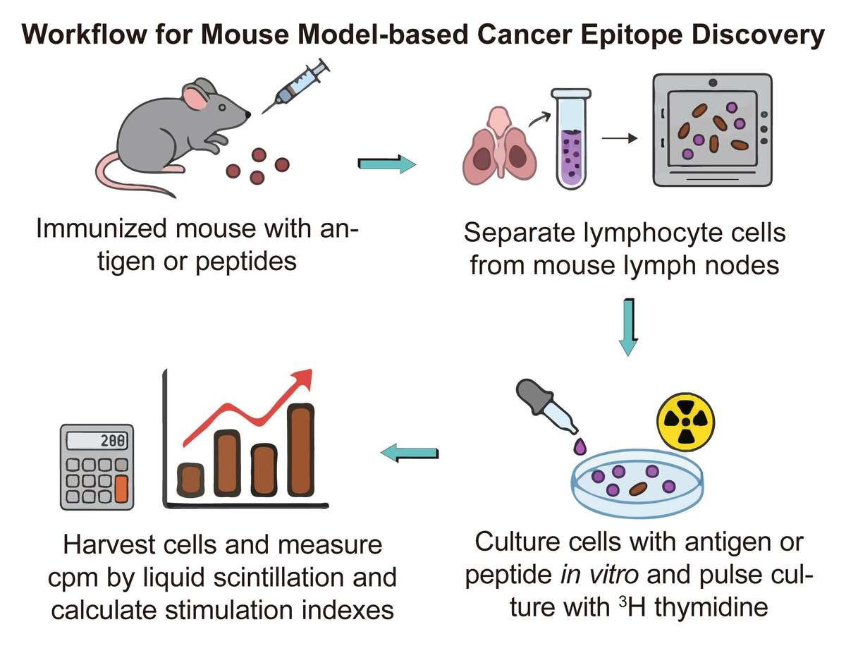
Workflow of Mouse Model-based Cancer Epitope Discovery Using 3H-thymidine Assay
Our workflow is designed for clarity and efficiency, ensuring a seamless experience from sample submission to final data delivery.

Why Choose Us?
In the fast-moving field of immuno-oncology, accurately assessing T cell function is essential. At Creative Biolabs, we provide robust, reliable solutions for measuring T cell proliferation—a critical step in developing new immunotherapies, vaccines, and drug candidates. As a trusted partner in immunogenicity studies, we combine deep scientific expertise with advanced technology to support your research goals. Our T cell-based 3H-thymidine incorporation assay remains a gold standard for evaluating immunogenicity, offering direct, reproducible measurement of T cell proliferation to help advance your most promising therapeutic programs.
Discover how we can help your project succeed. Request a consultation today to begin the conversation about your specific goals.
FAQs
Q1: How does your assay compare to colorimetric assays like MTT?
A1: Our T cell-based 3H-thymidine assay directly measures DNA synthesis, providing a more direct and reliable measure of proliferation compared to colorimetric assays, which measure metabolic activity and can be influenced by other factors.
Q2: Do you offer alternatives to the radioactive assay?
A2: While we specialize in the gold-standard 3H-thymidine assay for its precision, we can discuss your project requirements to determine if a non-radioactive method would be a suitable alternative.
Q3: How sensitive is the assay for low-frequency T cells?
A3: The 3H-thymidine assay is highly sensitive and can detect proliferation in low-frequency T cell populations, making it an excellent tool for epitope screening and rare T cell detection.
Customer Review
-
Direct Proliferation Measurement
Using Creative Biolabs' T cell-based 3H-thymidine assay in our research has significantly improved our ability to get a direct, quantitative measurement of antigen-specific T cell proliferation, which we couldn't achieve with alternative methods. – Ken*Bd
-
Superior to Colorimetric Assays
Creative Biolabs' 3H-thymidine assay provided us with reliable and consistent data, allowing us to confidently screen our library of peptides for a new vaccine. – Jn*Sh
Related Services
To further support your research, we recommend the following complementary services:
T and B Cell-based ELISPOT Assay
Creative Biolabs offers advanced ELISpot assays for cancer epitope analysis, leveraging its mature platform and expert team. This highly sensitive assay measures cytokine secretion activity to identify antigen-specific humoral and cellular immunity.
Learn More →
FluoroSpot Assay Service
Creative Biolabs offers the FluoroSpot assay, an advanced alternative to ELISpot. This highly sensitive assay uses fluorochrome-conjugated detection antibodies to detect secreted cytokines or antibodies, enabling functional analysis of up to seven subsets of immune cells.
Learn More →
Contact Us
Creative Biolabs is dedicated to accelerating your therapeutic research. Our team of experts is ready to discuss your specific needs and provide a tailored solution. For detailed information and to discuss your project, please reach out to us.
Reference
-
Ganesan, Nirosha, Steven Ronsmans, and Peter Hoet. "Methods to assess proliferation of stimulated human lymphocytes in vitro: a narrative review." Cells 12.3 (2023): 386. Distributed under Open Access license CC BY 4.0, without modification. https://doi.org/10.3390/cells12030386


 Fig.1 Schematic representation of 3H-thymidine assay.1

