Beyond single-analyte ELISPOT, we provide comprehensive services for analyzing a wide range of cytokine production. Our services also include immunogenicity assays to evaluate the safety and efficacy of therapeutic proteins.
Learn More →FluoroSpot Assay Service
Creative Biolabs is an industry leader in cellular immune monitoring, specializing in advanced FluoroSpot technology. With over two decades of experience, we help you streamline clinical trial processes and obtain high-quality, actionable data through our cutting-edge, multiplexed cellular analysis technology. Our service allows for a deeper understanding of immune responses with fewer samples, accelerating your research and development timelines.
Introduction What We Can Offer Workflow Why Creative Biolabs Customer Reviews FAQs Related Services Contact Us
Introduction of FluoroSpot Assay Service
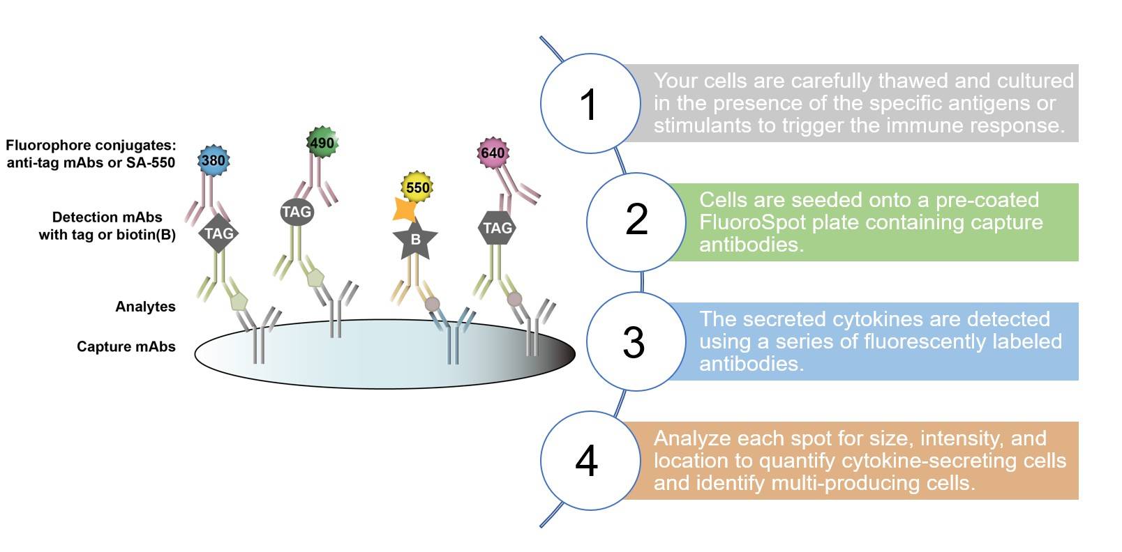
The FluoroSpot assay at Creative Biolabs has been developed as an alteration of colorimetric substrate-based traditional enzymatic ELISPOT detection systems. ELISPOT is a good choice for the functional analysis of single immune cells, while FluoroSpot can be used for the functional analysis of 7 subsets of immune cells. The FluoroSpot is capable of detecting secreted cytokines or antibodies with a high level of sensitivity, with the use of fluorochrome-conjugated detection antibodies.
To ascertain the potential of this service for your specific research, request a consultation.
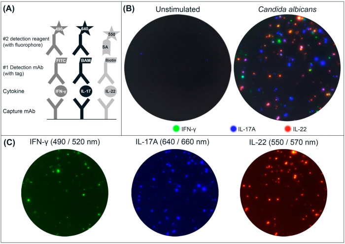 Fig.1 A triple FluoroSpot assay based on mAbs to human IFN-γ, IL-17A, and IL-22.1
What We Can Offer
| Common Cytokines Investigated | B cell FluoroSpot assays |
|---|---|
|
|
Let Creative Biolabs Help You with FluoroSpot Assay
Highlights
Superior Sensitivity
Our FluoroSpot assay is a highly effective tool with high sensitivity. It is an advanced version of ELISPOT that uses fluorescent dyes for detection, enabling it to detect even low frequencies of antigen-specific immune cells with remarkable precision and confidence.
Advanced Multiplexing
We enable the detection of two or more different cytokines simultaneously from the same sample. This powerful multiplexing capability provides a multi-parameter view of the immune response, allowing you to gain a deeper, more comprehensive understanding of cellular function.

Sample Efficiency
Our high-sensitivity assay requires a lower amount of peripheral blood mononuclear cells (PBMCs) to achieve a robust signal. This is critical for projects with limited patient material, ensuring you can still get comprehensive, actionable data.
Broad Applicability
The FluoroSpot assay can be used to evaluate both B-cell and T-cell responses in individuals. It is also an effective tool for clinical trial monitoring, helping to assess the efficacy and safety of new drug and vaccine candidates.
For a comprehensive review of our capabilities - Get a quote today.
Customer Reviews
-
Precise and Actionable Data
The precision and quality of the data we received from Creative Biolabs were exceptional. Their analysis of IFN-γ, IL-17A, and IL-22 responses from PBMCs was instrumental in our infectious disease research. The detailed report provided clear and actionable insights that directly supported our findings. - Dr. Rn, Immunology Group*. -
Streamlined Workflow
Creative Biolabs' service streamlined our workflow by eliminating the need for multiple single-analyte assays. We were able to get more data from a smaller sample size, which was crucial for our clinical trial with limited patient material. - Dr. Ms, Clinical Trials Team*.
FAQs
What types of samples can be used with the FluoroSpot assay Service?
Our service is optimized for use with single-cell suspensions like PBMCs and Tumor-infiltrating lymphocytes (TILs). If you have a different sample type, please contact us to discuss the feasibility and any required pre-processing steps.
How does the FluoroSpot assay compare to flow cytometry?
While flow cytometry provides a phenotypic analysis of cell populations, FluoroSpot offers a direct functional readout by quantifying the number and type of secreted molecules from a single cell. Both methods provide different but complementary data.
Related Services
ELISPOT Assay Service
Antibody Engineering
If your project requires custom antigens or antibodies, our expert team can develop and produce them. Our capabilities extend to antibody reformatting, humanization, affinity maturation, and immunogenicity prediction to help accelerate your therapeutic development pipeline.
Learn More →How to Contact Us
To fully leverage the capabilities of our cellular immunity research solutions, we invite you to contact our expert team. Scientists at Creative Biolabs have first-class technology and offer comprehensive service in applying the FluoroSpot assay. For more detailed information about our FluoroSpot service, please contact us immediately.
Reference
- Dillenbeck, Tomas et al. "Triple Cytokine FluoroSpot Analysis of Human Antigen-Specific IFN-γ, IL-17A and IL-22 Responses." Cells vol. 3,4 1116-30. 27 Nov. 2014. Distributed under an Open Access license CC BY 4.0, without modification. https://doi.org/10.3390/cells3041116
 Download our brochure
Loading case studies...
Online Inquiry