Cynomolgus Monkey Brain (NHP-TI126)
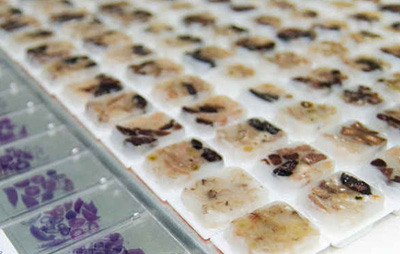
Non-Human Primate (NHP) Tissue Block Products, Cynomolgus Monkey,  Tissue Block
Category:Non-Human Primate (NHP) Tissue Block Products
Tag: Cynomolgus Monkey, Tissue Block
  • Please enter characters, no more than 250 characters; (0/250)
  • This tissue block from Creative Biolabs is isolated from Cynomolgus monkeys' brain. The sample has been verified as negative for multiple types of virus infections including Herpes-B Virus, SRV, SIV, and STLV-1, and can serve as a beneficial tool in a number of in vitro test and assay experiments such as PCR, WB, immunoprecipitation, immunofluorescent flow cytometry.

Detailed Product Description

  • Source: Healthy cynomolgus monkey
  • Applications: Given their robust resemblances to humans across physiological, behavioral, immunological, and genetic aspects, non-human primates serve as crucial models in a broad range of biomedical research, especially in bridging translational research from small animal models to humans. Creative Biolabs offers both standard formats and personalized non-human primate tissue preparations, catering to diverse in vitro testing applications.
  • System: Nervous System
  • Organ/Tissue: Brain
  • Shipping: Dry ice

Technical Specifications

  • Preservation Methods: Snap frozen
  • Quality Assurance: Tissue blocks are prepared by histologists with years of experience to be sure of excellent morphology and high quality.
  • Packaging: Securely packaged to maintain the tissue quality during shipping.
Downloadable Resources Published Data FAQs Customer Reviews
  • Cynomolgus Monkey Brain (NHP-TI126) Data Sheet: Detailed information.

This paper yields comprehensive research regarding the neurotoxic effects of dexmedetomidine compared to ketamine in the brains of fetal Cynomolgus monkeys. The study highlights the apprehension of anesthetics as a neurodegenerative effect on the developing brains, with special reference to the prospective neuroprotective properties of dexmedetomidine vs. anesthesia-induced neuronal injury. The histopathological examinations and NHP pharmacokinetic analysis, therefore, assessed neurotoxic effects and showed that, in a well-designed experimental setting, high doses of dexmedetomidine did not produce significant neuroapoptosis or neurodegeneration in the frontal cortex of the developing brain. This is an important finding that illustrates, in sharp contrast, the safety profile of dexmedetomidine versus the marked neurotoxicity seen with ketamine administration in the context of pediatric anesthesia. This study has the in-depth importance of guiding anesthetic practices in the safer development of neonatal and pediatric care. Therefore, it further illuminates the need for more research on anesthetic developmental neurotoxicity.

Activated caspase 3 (AC3) immunostained photomicrographs of the frontal cortex of fetal monkey brains of untreated control (A) and monkevs treated with ketamine (B). low-dose dexmedetomidine (C). and high-dose dexmedetomidine (D). Arrows in Fig.2B indicate AC3-positive cells.10X magnification.1Fig. 1. Activated caspase 3 (AC3) immunostained photomicrographs of the frontal cortex of fetal monkey brains of untreated control (A) and monkevs treated with ketamine (B). low-dose dexmedetomidine (C). and high-dose dexmedetomidine (D). Arrows in Fig.2B indicate AC3-positive cells.10X magnification.

Activated caspase 3 (AC3) immunostained photomicrographs of the frontal cortex of fetal monkey brains of untreated control(A) and monkeys treated with ketamine (B), low-dose dexmedetomidine (C), and high-dose dexmedetomidine. The AC3-positive cells shown in Fig. 2B can be clearly seen in Fig. 3B. 20X magnification.1Fig. 2. Activated caspase 3 (AC3) immunostained photomicrographs of the frontal cortex of fetal monkey brains of untreated control(A) and monkeys treated with ketamine (B), low-dose dexmedetomidine (C), and high-dose dexmedetomidine. The AC3-positive cells shown in Fig. 2B can be clearly seen in Fig. 3B. 20X magnification.

Immunohistochemical TUNEL-label photomicrograhs of the frontal cortex of fetal monkey brains of untreated control(A and monkeys treated with ketamine (B), low-dose dexmedetomidine (C), and high-dose dexmedetomidine (D). TUNEL. labeled cells as a result of ketamine treatment are clearly seen as brown cells in Fig. 4B. 20X magnification.1Fig. 3. Immunohistochemical TUNEL-label photomicrograhs of the frontal cortex of fetal monkey brains of untreated control(A and monkeys treated with ketamine (B), low-dose dexmedetomidine (C), and high-dose dexmedetomidine (D). TUNEL. labeled cells as a result of ketamine treatment are clearly seen as brown cells in Fig. 4B. 20X magnification.

Immunohistochemical TUNEL-label photomicrograhs of the frontal cortex of fetal monkey brains of untreated control(A and monkeys treated with ketamine (B), low-dose dexmedetomidine (C), and high-dose dexmedetomidine (D). TUNEL. labeled cells as a result of ketamine treatment are clearly seen as brown cells in Fig. 4B. 20X magnification.1Fig. 4. Immunohistochemical TUNEL-label photomicrograhs of the frontal cortex of fetal monkey brains of untreated control(A and monkeys treated with ketamine (B), low-dose dexmedetomidine (C), and high-dose dexmedetomidine (D). TUNEL. labeled cells as a result of ketamine treatment are clearly seen as brown cells in Fig. 4B. 20X magnification.

Reference

  1. Edward Koo., et al. " Neurotoxic effects of dexmedetomidine in fetal cynomolgus monkey brains." The Journal of Toxicological Sciences. 2014, 39: 251–262
  1. Q 1: How are the brain tissues ethically sourced from Cynomolgus monkeys?

    A: Therefore, procurement of these tissues should be from facilities that operate with great respect for animal welfare up to the observance of even high ethical standards.

  2. Q 2: What methods are used to ensure the high quality and preservation of brain tissues for research purposes?

    A: These include different methods of fixing, preserving, and storing tissues that help in maintaining cellular integrity so that the tissue can be put to histological examination.

  • High-Quality, Well-preserved,
    The use of Cynomolgus Monkey Brain samples from Creative Biolabs in our neurodegenerative research has provided us with a big leap. It includes samples that are high in quality, well-preserved, and key to back the study up with other than being ethically sourced.
  • Reliable and Reproducible
    The customer service quality of Creative Biolabs is really high, and they helped us in the proper selection of Cynomolgus Monkey Brain tissues for our project. The qualities of the tissues were excellent, and they assured us that our experimental results would be reliable and reproducible.

Click the button below to contact us or submit your feedback about this product.

Online Inquiry

This site is protected by reCAPTCHA and the Google Privacy Policy and Terms of Service apply.

NHP Biologicals from Creative Biolabs are for research, diagnostic, or manufacturing purposes only. They may not be intended for or are not approved for any other use.

USA
Tel:
Fax:
Email:
UK (for antibody and medium reagents only)
Tel:
Email:
Germany
Tel:
Email:

Enter your email here to subscribe.

Submit

Follow us on

Copyright © 2024 Creative Biolabs. All rights reserved.
Cart
  • 0
  • 0
Cart