Creative Biolabs provides services that include the most advanced TME-mimicking assays to assess the exhaustion status of immune cells, such as cytokine production, expression of inhibitory markers and immune cell degranulation. We offer multiple proprietary profiling methods that can help you to identify innovative strategies for reversing immune exhaustion and improving the efficacy of cancer immunotherapy.
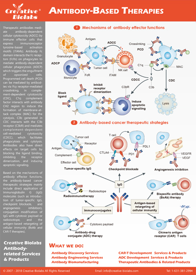
Background
T cell exhaustion is a sort of dysfunction or 'burnout' condition that often occurs in chronic infections and tumors and is associated with a loss of effector functions, including reduced cytokine secretion, proliferation, and cytotoxicity. A similar 'exhaustion-like' phenotype has been observed in NK cells that reside within the tumor microenvironment (TME). Dissecting these mechanisms is a critical area of research into how to improve immunotherapy, including adoptive transfer approaches, whether CAR-T therapy, TCR-T therapy, or NK cell therapy. Creative Biolabs focuses on addressing the mechanisms underlying both T cell and NK cell exhaustion through its specialized assays, helping researchers design more effective immunotherapies for solid and hematologic malignancies.
T Cell Exhaustion
T cell exhaustion is a state of functional decline that occurs due to persistent antigen exposure, such as in chronic infections and tumors. Exhausted T cells are characterized by:
Loss of cytokine production: Reduced levels of IL-2, IFN-γ, and TNF-α compromise immune function.
Upregulation of checkpoint markers: Proteins such as PD-1, CTLA-4, LAG-3, TIM-3, 2B4 / CD244 / SLAMF4, CD160, TIGIT are expressed in higher quantities, limiting T cell responses.
Epigenetic reprogramming: Exhausted T cells undergo chromatin remodeling, locking them in a dysfunctional state. Innovative strategies, including histone modifiers and checkpoint inhibitors, aim to reverse this state.
Fig.1 T Cell Exhaustion Markers.1,3
NK Cell Exhaustion
Natural killer (NK) cells exhibit exhaustion-like behavior within the tumor microenvironment (TME), characterized by:
Reduced cytotoxicity and IFN-γ production: Impaired ability to eliminate cancer cells.
Expression of inhibitory receptors: Increased levels of NKG2A, TIGIT, CD16, CD226, 2B4 and NCRs, hindering immune responses.
Partial responsiveness to checkpoint inhibitors: Although immune checkpoint inhibitors like PD-1 blockade have shown promise, the effects on NK cells are variable compared to T cells.
Fig.2 NK Cell Exhaustion.2,3
Key Technologies for T/NK Cell Exhaustion Assay
Technology Description
Common Targets
Examples of Detection
Flow Cytometry
Multiplex analysis of immune markers to detect exhaustion.
PD-1, TIM-3, LAG-3, TIGIT
Quantifies checkpoint molecules on CAR-T cells.
Western Blotting
Simultaneous measurement of cytokines and intracellular proteins.
IFN-γ, IL-2, TNF-α
Detects cytokine expression levels in T cells.
Cytokine Profiling
Measures production of multiple cytokines simultaneously.
IL-2, IFN-γ, CCL5
Reveals immune cell dysfunction.
Single-Cell RNA Sequencing
(Recommended) Offers molecular profiling of individual cells.
Exhaustion-related genes: NR4A, BATF3
Identifies transcriptional signatures of exhausted T/NK cells.
Degranulation Assay
Assesses functional cytotoxicity.
Granzyme B, Perforin
Detects effector functions of NK cells.
T/NK Cell Exhaustion Assay Workflow
In this assay, fresh isolated human PBMCs are stimulated with a fixed concentration of compounds for three days. The media is harvested for hallmark measurements. These assays enable precise measurement of cell dysfunction, helping researchers optimize therapies such as CAR-T cell treatments and checkpoint inhibitors.
Isolation of PBMCs: Peripheral blood mononuclear cells are isolated as the starting material.
Stimulation Protocol: Cells are stimulated with a defined concentration of immune-modulating compounds over three days.
Measurement of Key Markers: Exhaustion markers (e.g., PD-1, CTLA-4) and cytokine production are analyzed using advanced technologies mentioned above.
Data Interpretation: Customized reports provide actionable insights on immune exhaustion and therapeutic targets.
Fig.3 Workflow of T/NK Cell Exhaustion Assay.
Applications of T/NK Cell Exhaustion Assay
Application Description
Use Cases
CAR-T Therapy Optimization
Identifies exhaustion markers during CAR-T cell production and persistence.
Enables refinement of CAR-T manufacturing processes.
Checkpoint Blockade Evaluation
Assesses effects of inhibitors like PD-1 and CTLA-4 on reversing exhaustion.
Optimizes combination therapies with checkpoint inhibitors.
Preclinical Drug Screening
Tests new compounds targeting exhaustion pathways.
Facilitates drug development for enhancing immune therapies.
Identifies exhaustion-related markers predictive of treatment outcomes.
Helps guide personalized therapy decisions.
Immune cell exhaustion assays from Creative Biolabs are state-of-the-art tools that help to research and unleash the true potential of TCR-T, CAR-T and NK cell therapy. Our assays enable researchers to improve immunotherapy protocols by identifying exhausted immune cells through advanced flow cytometry, cytokine profiling and data interpretation. Besides, Creative Biolabs' solutions have generated significant interest in immune cell research, accompanied by the growing need of effective cancer treatments. We are now working side by side with leading scientists in the field of adoptive cell transfer and breakthrough checkpoint blockade therapies. Our efforts allow us to expand our services to meet the demands of academic researchers as well as the pharmaceutical industry, to ensure that CAR-T and NK cell therapies are as impactful as possible in the clinic. For more details, please feel free to contact us for project quotations and more detailed information.
References
Okoye, Isobel S., et al. "Coinhibitory receptor expression and immune checkpoint blockade: maintaining a balance in CD8+ T cell responses to chronic viral infections and cancer." Frontiers in immunology 8 (2017): 1215.
Bi, Jiacheng, and Zhigang Tian. "NK cell exhaustion." Frontiers in immunology 8 (2017): 760.
Distributed under Open Access license CC BY 4.0, without modification.
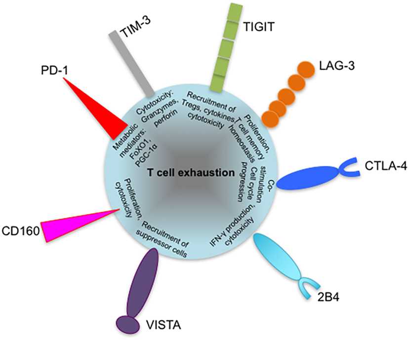 Fig.1 T Cell Exhaustion Markers.1,3
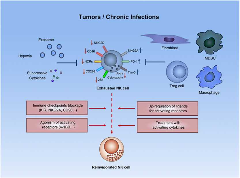 Fig.2 NK Cell Exhaustion.2,3
 Fig.3 Workflow of T/NK Cell Exhaustion Assay.
 TCR/CAR Expression Analysis
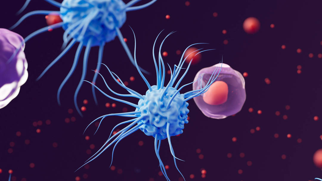
 Download our brochure