Single Domain Antibody (SdAb) Discovery Service for In Vivo Functional Molecular Imaging
Molecular imaging is a non-invasive technique employed to visualize, characterize, and quantify biological processes at the molecular and cellular levels within humans or other living organisms. In essence, injecting and following molecular imaging tracer, which comprises a targeting vehicle joined to a detection label, is a convenient functional molecular imaging approach. In recent years, various molecular imaging tracers have been developed for (pre)clinical research fields such as oncology, cardiology, neurology, or rheumatology. Creative Biolabs offers novel single domain antibody (sdAb) discovery services to generate high-specific diagnostic reagents for in vivo functional molecular imaging purposes.
SdAb as In Vivo Molecular Imaging Tracer
The rapidly growing field of in vivo molecular imaging has inspired the development of many imaging tracers, each with its advantages and limitations. In this area, sdAbs match the requirements of the ideal molecular imaging tracer because of their unique properties:
sdAbs bind tightly to the targets and have affinities in the low nanomolar to high picomolar range.
sdAbs are highly stable to tolerate numerous labeling strategies (e.g., rationale, near-infrared fluorophores, and fluorescent proteins) to meet the various in vivo imaging requirements modalities.
sdAbs can rapidly distribute through the bloodstream, reaching tissues homogenously.
sdAbs easily penetrate tissues, also accessing cryptic antigens (e.g., located behind the blood-brain barrier).
sdAbs from camelid share a higher degree of sequence identity with human VHs and have a low immunogenic potential.
sdAbs are cleared from blood rather quickly when unbound due to the small molecular weight, resulting in a contrast-enhanced imaging signal and reduced accumulation of labeled fragments in the liver, lowering radiation burden.
Collectively, these attributes render sdAbs highly suitable for in vivo molecular imaging probes in both preclinical and clinical settings. Nonetheless, a significant limitation of sdAb-based imaging is their high non-specific uptake in the kidneys and bladder. Potential solutions to this issue include the development of multivalent sdAbs or sdAb-Fc fusion constructs. The fusion of sdAbs with Fc fragments appears particularly promising due to this option can also extend half-life and enhance tumor penetration capabilities.
Fig. 1 sdAb-based in vivo molecular imaging.1
In the evolving landscape of integrated diagnosis and treatment, sdAbs are poised to play pivotal roles. They are instrumental in tumor diagnosis, assessment, and prediction prior to initiating therapeutic protocols, as well as in dynamic monitoring during treatment to detect potential disease recurrence. To date, various diagnostic imaging techniques have been developed and employed using sdAbs as in vivo tracers. These include nuclear imaging modalities such as positron emission tomography (PET) and single-photon emission computed tomography (SPECT) with radionuclides like 99mTc, 89Zr, or 68Ga; optical imaging with near-infrared (NIR) fluorescent dyes; ultrasound imaging using microbubbles; computed tomography (CT); and magnetic resonance imaging (MRI).
SdAb-based Tumor Imaging
Tumor imaging represents the most extensively researched application of sdAb-based molecular imaging. sdAbs targeted against tumor-associated proteins and labeled with a radionuclide have been successfully applied as tracers for noninvasive in vivo tumor imaging via PET and SPECT. In the field of oncology, single domain antibodies (sdAbs) have been engineered for non-invasive preclinical screening of cancers, especially those targeting EGFR1, HGF, HER2, prostate-specific membrane antigen (PSMA), CEA, and CIAX. These sdAbs are also utilized for monitoring cancer therapy through SPECT/CT or PET/CT imaging modalities.
SdAb-based Optical Imaging
Compared to SPECT and PET in which approximately 1 h is required for one scan, sdAb-mediated optical imaging with NIR takes a substantially short period (less than 1 min per image, with preclinical imaging systems). Besides, due to the readily available conjugation technology, the use of sdAbs in optical imaging for image-guided surgery is highly promising. Considering all these aspects, the sdAb-based platform for optical molecular imaging has rationally set up a new benchmark of rapid optical imaging.
SdAb-based Ultrasound Imaging
The utilization of sdAb-targeted microbubbles offers a safe, rapid, and cost-effective scanning method, delivering relatively high-quality images. For instance, VCAM1-specific sdAbs conjugate to lipid microbubbles can be used as contrast agents for ultrasound imaging, which contribute to evaluating potential adhesion sites for melanoma cell extravasation and metastasis.
Functional molecular imaging techniques, widely utilized in clinical settings, enable the non-invasive quantification and visualization of various diseases in vivo. As promising, small-sized, high-affinity imaging tracers, sdAbs facilitate effective tissue penetration and early acquisition of high-quality images, offering a comprehensive disease evaluation and supporting personalized precision therapy. Creative Biolabs, actively engaged in the development and production of novel sdAbs, can assist in creating specific sdAbs tailored to your functional molecular imaging needs. For further information about our services, please feel free to contact us.
Reference
Debie, Pieterjan, Nick Devoogdt, and Sophie Hernot. "Targeted nanobody-based molecular tracers for nuclear imaging and image-guided surgery." Antibodies 8.1 (2019): 12. Distribute under Open Access license CC BY 4.0, without modification.
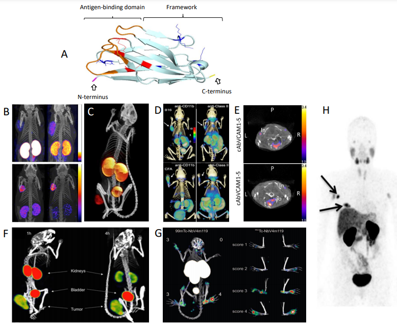 Fig. 1 sdAb-based in vivo molecular imaging.1