As a great breakthrough in stem cell reprogramming field, induced pluripotent stem cells (iPSCs) have been worldwide used in genetic modifications and cell therapy field. iPSCs provide great hope to patients and scientists because of their potential to differentiate into any cell types to cure diseases, reverse injuries and offer highly targeted therapies to improve health outcomes. iPSCs have generated a lot of interest and research into their potential in restoring vision. With years of experiences and advanced technologies, Creative Biolabs is recognized as the world leader for providing the most diverse portfolio of iPSCs differentiation studies. Now, we apply our expertise in the iPSC-derived ocular differentiation service to our worldwide customers.
iPSCs are derived from somatic cells that have been reprogrammed back into embryonic-like pluripotent stem cells which are capable of unlimited, undifferentiated proliferation in vitro and still maintain the capacity to differentiated into a wide range of somatic cells needed for therapeutic purposes. The most promising feature of iPSCs in cell therapy is that they can avoid the post-transplant immune suppression because of the direct origination from the transplant recipient. iPSCs differentiation to specialized cells needs professional guidance and expertly optimized growth media for their successful culture and propagation. The efficient differentiation of human iPSCs into functional relevant cell types is an important prerequisite for the successful development of iPSC-based treatment and modeling strategies. iPSC has the capacity to differentiate into functional cells of mesodermal lineage, including neural and retina cells, which has great potency for treatment of ocular disorders.
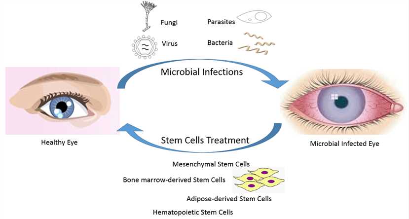
 Fig.1 Treatment of ocular microbial infections by iPSCs.1
Creative Biolabs offers a wide range of custom iPSC-derived ocular differentiation products and services for our clients all over the world.
We offer a comprehensive range of iPSC-derived differentiated ocular cells, including retinal precursor cells, retinal ganglion cells, cone and rod photoreceptor cell and so on, along with specialized growth media. The origination of our provided iPSCs comes from healthy donors and patients of specific disease backgrounds. We can also reprogramme the somatic cells you specially provided.
We provide cost-effective services to training and guide investigators in the creation of ocular- and retinal- specific cells from your iPSCs. Our expertise focuses on the reprogramming cells to iPSCs and differentiating them to various ocular cell types. The protocols are fully tailored to achieve both a high differentiation efficiency, and level of purity whilst retaining minimal lot-to-lot variation.
We can offer a comprehensive series of products to support iPSCs differentiation. These differentiated cells provide a highly desirable in vitro platform for high content toxicity and drug screening and as a feasible alternative to animal and embryonic stem cell models. Our kits maximize workflow efficiency and are applicable for drug toxicity and small molecule screening. It consists of a set of supplements that enable efficient differentiation of human pluripotent stem cells to ocular cells.
Creative Biolabs has successfully completed numerous iPSC-derived ocular differentiation projects. It is excited that our iPSC-derived cell models have been widely used for proof of concept studies and drug screening for new therapeutics. We are happy to offer our off-the-shelf product portfolio and outsourced services to help you get landmark development. If you have any special needs in iPS cell differentiation, please contact us. Our cost-effective services will be tailored to meet your experimental requirements.
Below are the findings presented in the article related to ocular cell differentiation from iPSC.
1. Researchers have used the Tie2 - GFP mouse model to generate two independent iPSC lines and, using these cell lines, developed an effective differentiation protocol capable of generating choroidal endothelial cells (CECs), which can be used for future disease modeling and cell replacement studies.
They characterized iPSC-derived CEC. RT-PCR analysis showed that iPSC-CEC expressed CEC-specific markers CA4, FOXA2, and PLVAP. iPSC-CEC lines derived from Tie2 - GFP were the most similar to primary CEC compared to other commercially available EC lines.
2. Another research team has utilized a commercially available automated cell culture platform to culture and differentiate hiPSC in a reproducible and scalable process to generate patient-derived retinal pigment epithelial (RPE) cells for downstream use, including drug testing.
They primarily used hiPSC from multiple donors suffering from age-related macular degeneration (AMD) to differentiate into RPE, and subsequently measured mitochondrial function after exposure of donor hiPSC-RPE lines to compounds that affect mitochondrial metabolism. One of the donor hiPSC-RPE lines was tested as follows.
Reference
For Research Use Only. Not For Clinical Use.