
Species-Relevant T Cell Activation Evaluation for Veterinary Antibody Programs
T-cell activation is a context-dependent process shaped by antigen recognition, co-stimulation, receptor engagement, cell source, antibody presentation, and assay timing. Creative Biolabs develops canine and feline assays to characterize whether a therapeutic antibody produces an intended immunomodulatory effect or an unwanted cellular response relevant to early immunogenicity and safety assessment.
Studies can use PBMCs, enriched T cells, or APC–T-cell co-cultures with mechanism-matched positive, negative, and isotype controls. Direct activation phenotyping is integrated with proliferation and selected cytokine measurements where appropriate, supporting candidate comparison without treating any single in vitro endpoint as a standalone predictor of anti-drug antibody formation or clinical immunogenicity.
T Cell Activation Evaluation for Immunogenicity Risk and Immune Modulation
We first define whether the study is intended to investigate unwanted immunogenicity-related T-cell responses, intended pharmacologic immune modulation, or both. This distinction determines the most appropriate cell system, stimulus, controls, and interpretation.
Immunogenicity-Oriented T Cell Response Assessment
Evaluate candidate-associated T-cell activation or proliferation using species-relevant primary immune cells. Whole antibody, defined peptide material, or APC-mediated presentation formats can be selected according to the study question and assay feasibility.
Direct Antibody-Mediated T Cell Modulation
Assess agonistic, blocking, checkpoint, or other T-cell-directed mechanisms in a controlled stimulation system when the antibody itself is expected to alter T-cell activation as part of its pharmacology.
APC–T Cell Co-Culture Evaluation
Use antigen-presenting cells and autologous or compatible T cells when uptake, processing, presentation, co-stimulation, or cell-cell interaction is important to the biological question.
Dose-Response, Time-Course, and Donor Profiling
Compare concentrations, sampling windows, and multiple veterinary donors to identify response patterns and biological variability that may be missed by a single-condition experiment.
Canine and Feline Immune Cell Systems for T Cell Activation Evaluation
The cell format is selected according to whether the study needs preserved immune-cell interactions, a more direct T-cell readout, or an antigen-presentation context relevant to immunogenicity-oriented assessment.
PBMC-Based T Cell Activation Assay
Preserve interactions among T cells, monocytes, antigen-presenting cells, and other immune populations while evaluating candidate-associated activation, proliferation, and complementary cytokine responses.
Enriched T Cell Assay
Reduce confounding from non-T-cell populations when the question focuses on direct receptor engagement, activation phenotype, proliferation, or intrinsic T-cell responsiveness.
Antigen Presentation and Co-Stimulation Context
Configure APC-containing cultures, peptide or protein stimulation, anti-CD3/co-stimulatory controls, and other assay conditions needed to distinguish antigen-specific responses from nonspecific immune activation.
Veterinary Donor Panel Strategy
Where sample availability permits, evaluate multiple healthy canine or feline donors to capture biological response variability and support comparative candidate assessment.
Integrating Activation Markers, Proliferation, and Complementary Cytokine Readouts
T-cell activation, proliferation, and cytokine output describe related but non-equivalent biology. We combine orthogonal endpoints so a candidate is not classified from one marker alone, particularly when the data will contribute to immunogenicity-oriented risk assessment.
| Assay Question | Example Readouts | What the Readout Supports | Interpretation Boundary |
|---|---|---|---|
| Early activation phenotype | Flow cytometric CD25, CD69, or other validated species-relevant activation markers | Evidence that defined T-cell populations change activation phenotype after candidate exposure or controlled stimulation | Marker upregulation depends on timing, baseline expression, gating strategy, and T-cell subset context |
| T cell proliferation | Dye dilution, DNA-synthesis methods, direct cell counting, or another validated proliferation approach | Evidence of sustained cell-division responses that complement early activation-marker measurements | Proliferation is downstream of activation and can be affected by viability, cytokines, and culture conditions |
| Complementary cytokine response | Selected IL-2, IFN-γ, TNF-α, or other mechanism-relevant cytokines | Additional evidence about functional immune signaling associated with the observed T-cell response | Cytokine output is not treated as a standalone predictor of ADA formation or clinical immunogenicity |
| T cell subset response | CD4/CD8 phenotyping, activation-marker co-expression, or other validated subset markers | Determines whether the response is concentrated within a defined T-cell compartment | Panel design depends on species-specific reagent availability, marker biology, and validation |
| Viability and nonspecific activation control | Viability dyes, unstimulated cells, isotype controls, and mechanism-matched positive controls | Helps distinguish meaningful T-cell responses from toxicity, background, or generalized assay stimulation | Control performance is required before interpreting candidate-associated differences |
Separate Pharmacology from Immunogenicity-Oriented Signals
If the antibody target directly regulates T cells, activation may reflect intended pharmacology rather than antigen-specific immunogenicity. Mechanism-matched controls are therefore built into interpretation.
T Cell Data Are One Component of Immunogenicity Risk
In vitro T-cell responses support comparative risk assessment but do not independently predict ADA incidence, which also depends on product, exposure, and host factors.
Five-Stage Veterinary Antibody T Cell Activation Evaluation Workflow
The five-stage workflow moves from project planning and immune-cell preparation through assay execution, multi-parameter analysis, and decision-oriented reporting.
Recommended Starting Information
- Antibody target and proposed mechanism
- Canine or feline species context
- Antibody format, concentration, and available material
- Known agonistic, blocking, or checkpoint activity
- Preferred cell source or available donor material
- Required comparison candidates and decision point
Typical Deliverables
- Assay plan and optimized test conditions
- Flow-cytometry gating and activation-marker data
- Proliferation and selected cytokine datasets
- Dose-response and time-course comparisons
- Control-normalized candidate comparison
- Methods, analyzed results, and interpretation report
Need to distinguish intended T-cell pharmacology from an unwanted immune response?
Share your antibody target, species, intended mechanism, and available material. We can configure a focused T-cell assay with controls that support both functional interpretation and immunogenicity-oriented risk assessment.
Published Data on Canine T Cell Activation Assay Design
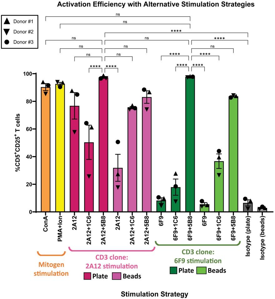
Davis et al. compared multiple canine T-cell stimulation strategies using PBMCs and quantified activation after three days by CD25 expression on CD5+ T cells. Their data showed that stimulation format and the selected αCD3/αCD28 combination materially affected the observed activation response, with plate-bound αCD3 combined with the 5B8 αCD28 co-stimulatory antibody among the stronger antibody-based conditions.1
The study illustrates why veterinary T-cell assays require species-relevant reagents, validated stimulation conditions, appropriate controls, and clearly defined activation endpoints. Creative Biolabs applies this logic to canine and feline antibody evaluation by integrating donor-aware cell systems, activation phenotyping, proliferation, and complementary functional readouts. When the goal is immunogenicity assessment, results are interpreted as comparative cellular risk evidence rather than as a standalone prediction of future ADA formation.
Why Choose Creative Biolabs for Veterinary T Cell Activation Evaluation?
Assay design is built around the antibody mechanism and the biological question so activation evidence remains interpretable across canine and feline programs.
Species-Relevant Assays
Canine and feline cell systems selected for the intended antibody mechanism.
Orthogonal Readouts
Activation markers, proliferation, and functional signals support integrated interpretation.
Mechanism-Matched Controls
Controls distinguish antibody effects from baseline stimulation and assay background.
Decision-Oriented Reporting
Comparative datasets support candidate selection and downstream assay planning.
Veterinary Antibody T Cell Activation Evaluation FAQs
References
- Davis, Treyvon W., et al. "Optimizing canine T cell activation, expansion, and transduction." PLOS ONE 20.9 (2025): e0324403. https://doi.org/10.1371/journal.pone.0324403.
- Distributed under Open Access license CC BY 4.0, without modification.