
Assess Veterinary Antibody Immunogenicity Risk Before Resource-Intensive Studies
Unwanted immune recognition can alter antibody exposure, reduce pharmacologic activity, or complicate interpretation of later studies. Creative Biolabs develops in vitro immunogenicity evaluation plans around the intended canine or feline species, antibody format, molecular characteristics, available donor cells, and the specific comparison required at the current development stage.
Rather than treating one assay as a direct predictor of anti-drug antibody formation, we combine species-relevant cellular readouts with appropriate controls and donor-aware interpretation. The resulting data are used as comparative risk indicators for candidate ranking, engineering decisions, assay refinement, and planning of subsequent in vivo immunogenicity or broader preclinical evaluation.
Antibody Immunogenicity Evaluation Services
Unwanted immune recognition is an important development risk for antibody-based therapies in companion animals because it can reduce therapeutic exposure, diminish activity, or complicate tolerability and downstream study interpretation. Creative Biolabs provides early-stage in vitro immunogenicity evaluation designed around the target canine or feline immune system. By combining species-relevant immune cells with fit-for-purpose cellular readouts, we support comparative candidate assessment, deimmunization or engineering decisions, and rational planning before more resource-intensive in vivo studies.
T Cell Activation Evaluation
T helper-cell activation is a key initiating event in many adaptive immune responses to therapeutic proteins. Using PBMCs or other appropriate immune-cell preparations from healthy, treatment-naïve canine or feline donors, we assess candidate-associated T-cell responses with project-matched activation or proliferation readouts. The resulting donor-aware data help identify relative cellular-response risk and support comparison of antibody candidates or engineered variants.
Cytokine Response Profiling for Immunogenicity Risk Assessment
Cytokine measurements can provide a complementary readout alongside T-cell activation or proliferation when evaluating candidate-associated immune responses. We measure project-relevant cytokines using sensitive plate-based, multiplex, ELISpot, intracellular, or other fit-for-purpose approaches as appropriate. Because cytokine release can also reflect innate immune activation or acute infusion-related mechanisms, these data are interpreted together with donor context, controls, material quality, and T-cell findings rather than as an independent surrogate for future ADA formation.
Veterinary Antibody In Vitro Immunogenicity Evaluation Workflow
The workflow follows four connected stages from project definition and species-relevant immune-cell sourcing through assay execution and comparative interpretation.
- Optional, project-dependent APC processing and presentation assays where suitable reagents and biology support the model
- T-cell activation or proliferation assays
- Complementary cytokine-response profiling with project-relevant analytes
Recommended Starting Information
- Target species and intended indication
- Antibody sequence, isotype, and format
- Purity, aggregation, and material history
- Available canine or feline donor cells
- Existing binding or functional data
- Comparator, engineering, or ranking objective
Typical Final Deliverables
- Customized assay plan and controls
- Raw and processed cellular-response data
- Donor-level and group-level comparisons
- T-cell and cytokine response summaries
- Relative immunogenicity-risk interpretation
- Consolidated methods and next-step recommendations
Match the Immunogenicity Question to the Right Cellular Readout
A useful in vitro immunogenicity study begins with a defined comparison. The same readout should not be interpreted identically across candidate ranking, sequence engineering, cytokine-response investigation, and donor-variability questions.
| Evaluation Question | Representative Approach | Important Controls / Variables | Decision-Oriented Output |
|---|---|---|---|
| T-Cell Response | PBMC or T-cell proliferation, activation-marker analysis, or complementary T-cell functional readouts. | Untreated cells, positive stimulation control, vehicle or formulation control, donor identity, concentration, and exposure time. | Candidate-associated response frequency, magnitude, and comparative rank across donors. |
| Cytokine Response | ELISA, ELISpot, multiplex immunoassay, or intracellular cytokine analysis used as a complementary readout alongside T-cell activation or proliferation. | Baseline cytokine release, assay sensitivity, cell composition, endotoxin, aggregates, and positive controls. | Contextual cytokine-response profile that supports interpretation of candidate-associated cellular immune responses. |
| Optional APC / Antigen Processing and Presentation | Project-dependent evaluation of antibody uptake and processing, APC activation, peptide-presentation-related responses, or APC–T-cell co-culture where suitable reagents and biology support the model. | APC phenotype and maturation state, antibody concentration, processing conditions, peptide-presentation context, donor source, and matched controls. | Upstream mechanistic evidence relevant to MHC II-dependent CD4+ T-cell recognition; interpreted as a supporting rather than standalone immunogenicity endpoint. |
| Variant Comparison | Side-by-side testing of parental and engineered sequences, formats, stressed material, or selected lots. | Matched concentration, quality attributes, formulation, donor panel, assay day, and acceptance criteria. | Relative risk ranking and evidence for whether a modification changed the cellular-response profile. |
What the Data Can Support
- Relative comparison of candidates tested under the same experimental framework.
- Identification of donor-dependent response patterns and assay-sensitive candidates.
- Selection of candidates or variants for additional characterization or engineering.
- Rational planning of subsequent in vivo immunogenicity or broader preclinical studies.
What Requires Additional Evidence
- Actual anti-drug antibody incidence after administration in the target population.
- Clinical consequences for exposure, efficacy, hypersensitivity, or tolerability.
- Population-wide effects not represented by the donor panel or assay conditions.
- Causal attribution when material quality, endotoxin, aggregation, or Fc-mediated effects may contribute.
Need a Focused Immunogenicity Pilot?
Share your species, antibody format, material quality information, comparator strategy, and current data. We can recommend a focused cellular assay or a multi-readout evaluation plan.
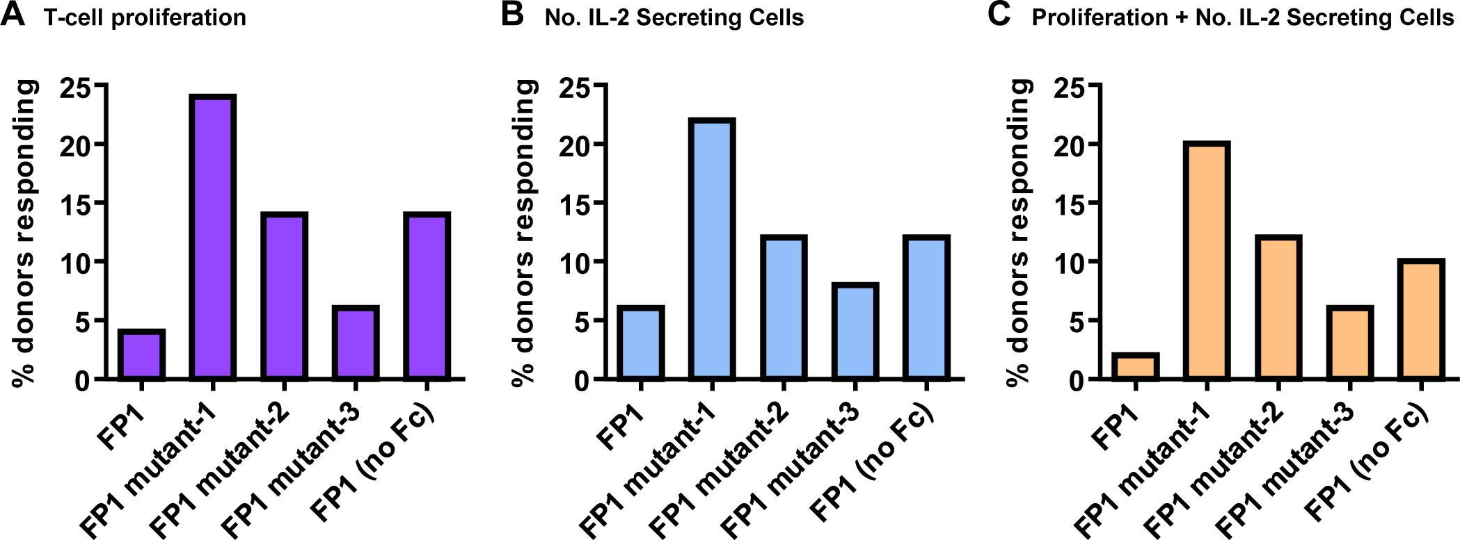
Published Data: Comparative T-Cell Responses Can Distinguish Closely Related Biotherapeutic Variants
Joubert et al. evaluated an in vitro comparative immunogenicity assessment using PBMCs from healthy human donors and showed that closely related biotherapeutic variants differing by one or two amino acids produced distinguishable T-cell proliferation and IL-2 ELISpot response patterns. The study supports the value of donor-based cellular assays for relative immunogenicity-risk comparison, while the human system should not be interpreted as direct validation of canine or feline clinical immunogenicity.
The comparative experimental framework is conceptually relevant to veterinary antibody evaluation when adapted to species-relevant immune-cell systems: matched candidates can be compared under controlled conditions using proliferation, cytokine-secreting-cell, and multi-donor readouts, with material quality and sequence differences treated as potential contributors to response. Creative Biolabs applies this comparative principle using canine or feline immune-cell systems and project-specific controls to support lead ranking, engineering follow-up, and downstream study design.
Advantages of Veterinary Antibody In Vitro Immunogenicity Evaluation
The service is designed around species relevance, assay fit, comparative interpretation, and transparent reporting rather than overextending one cellular readout into a clinical prediction.
Species-Relevant Design
Canine or feline immune-cell context guides model, donor, control, and readout selection.
Multi-Readout Evaluation
T-cell and cytokine endpoints provide complementary evidence for comparative candidate assessment.
Decision-Focused Comparison
Matched assay conditions support clearer ranking of candidates, variants, formats, or selected lots.
Transparent Interpretation
Reports distinguish measured cellular signals from conclusions requiring later in vivo evidence.
Veterinary Antibody In Vitro Immunogenicity Evaluation FAQs
References
- Joubert, Marisa K., et al. "Use of In Vitro Assays to Assess Immunogenicity Risk of Antibody-Based Biotherapeutics." PLOS ONE 11.8 (2016): e0159328. https://doi.org/10.1371/journal.pone.0159328
- Distributed under Open Access license CC BY 4.0, without modification.