Creative Biolabs is offering the most comprehensive services for antibody development projects. With strict regulation and effective execution, we are dedicated to providing the most valuable solutions to complete your projects.
HighlightsWith over a decade of experience in phage display technology, Creative Biolabs can provide a series of antibody or peptide libraries that are available for licensing or direct screening. These ready-to-use libraries are invaluable resources for isolating target-specific binders for various research, diagnostic or therapeutic applications.
HighlightsCreative Biolabs has established a broad range of platforms for developing novel antibodies or equivalents. These cutting-edge technologies enable our scientists to meet your demands from different aspects and tailor the most appropriate solution that contributes to the success of your projects.
HighlightsWith deep understanding in antibody-related realms and extensive project experience, Creative Biolabs offers a variety of references to help you learn more about our capacities and achievements, including infographic, flyer, case study, peer-reviewed publications, and all kinds of knowledge that can assist your projects. You are also welcome to contact us directly for more specific solutions.
HighlightsGet a real taste of Creative Biolabs, one of the most professional custom service providers in the world. We are committed to providing highly customized comprehensive solutions with the best quality to advance your projects.
HighlightsDendritic cells (DCs) are the major population of antigen-presenting cells (APCs) in vivo, pivotal for activating and stimulating CD4+ T cells to differentiate into effective T cells. This activation process involves the interaction between peptide-MHCII complexes and T cell receptors to constitute part of costimulatory signals. Notably, conventional dendritic cells 2 (cDC2) exhibit significantly greater antigen uptake and presentation of processed antigens on MHCII compared to conventional dendritic cells 1 (cDC1). Consequently, cDC2 induce a much higher proliferation of CD4+ T cells. These differences are attributed to the expression of IRF4 in cDC2, which enhances peptide-MHCII complex formation within the cells. In addition to activation and proliferation signals, the interaction between DCs and with CD4+ T cells during antigen presentation determines the differentiation of Th cells.
Ex vivo immunogenicity assessment is an essential component of preclinical testing for biological medicines prior to clinical trials. The purpose of this testing is to evaluate the potential immunogenicity of all new therapeutic products, including biological and biosimilar products, ensuring their safety before advancing to clinical phases.
Furthermore, this assessment aids in the study of naive T cell proliferation and DC-T cell proliferation, which are indispensable for understanding how the immune system is activated and responds to threats. Therefore, this methodology occupies a central position in immunology research, offering a versatile tool for analyzing the intricate complexity of the immune system.
Dendritic cells, renowned for their potent antigen presentation capabilities, play a pivotal role in initiating specific responses in T cells. They activate naive T cells and enhance the expression of T-cell receptor ligands and co-stimulatory molecules. In addition, DCs also provide crucial differentiation signals for CD4+ T cells.
For example, in the immunogenicity evaluation of antibody drugs, antibodies are exogenous antigens, so the body generally produces an immune response through the class II HLA pathway. Dendritic cells first capture these antigens through processes such as phagocytosis, endocytosis, or micropinocytosis of exogenous antibodies during antigen presentation. Subsequently, these DCs undergo maturation and migrate to the lymph nodes. Next, the exogenous antigen undergoes degradation into segments, forming peptide-MHC complexes with antigen peptides via major histocompatibility complex II (MHC-II) molecules. The mature antigenic peptide-MHC complexes are then presented on the surface of dendritic cells.
T cell activation follows a dual signal recognition mechanism, namely the recognition of antigenic peptide-MHC complexes and TCR, as well as the recognition of co-stimulatory molecules. Furthermore, the influence of cytokines serves as a third signal that promotes the proliferation and differentiation of T cells.
The initial interaction occurs between the T cell receptor and its ligand. Antigens, foreign substances invading the body, are recognized by the immune system, triggering an immune response. Upon entering the body, antigens are first captured by dendritic cells, specialized antigen-presenting cells that process them into small peptide segments recognizable by T cell receptors. These treated peptides then bind to MHC Class II molecules on dendritic cells. MHC Class II molecules act as proteins on the surface of antigen-presenting cells, forming a miniature "showcase" capable of hosting processed peptides. Loaded with small peptides, MHC Class II molecules are displayed on the surface of dendritic cells, simultaneously recognized by T cell receptors. This process is known as the "first signal," critical for initiating the T cell immune response. In this way, although antigens are thus "presented" to naive T cells, this alone does not activate them.
Activation of naive T cells necessitates a second signal, provided by co-stimulatory molecules. These specific proteins on the surface of dendritic cells bind to receptors on naive T cells, supplying the essential "second signal" to initiate the immune response. Co-stimulatory molecules include the oligonucleotide-induced B7 molecules (e.g., CD80 and CD86) and the ICOS ligand. Such co-stimulation can further amplify the T cell's response to the antigen.
Upon receiving these dual signals, T cells undergo activation and initiate proliferation. Consequently, copious amounts of T cells are generated in the body, poised to target antigens and combat pathogens. Furthermore, the interaction between dendritic cells and CD4+ T cells, while pivotal in determining the fate of T cell differentiation through antigen presentation, extends beyond mere promotion of CD4+ T cell activation and proliferation. The intricate mechanisms of cell-cell interaction during antigen presentation play a significant and indispensable role in regulating immune responses.
Overall, this process ensures that T cells selectively respond to external pathogens without targeting the body's own tissues.
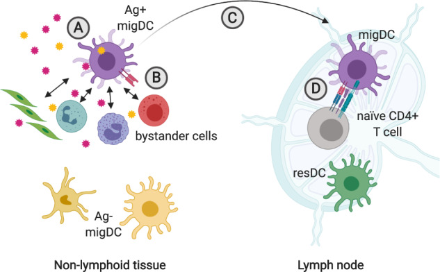 Fig. 1 Migrating dendritic cells integrate signals from both antigens and adjacent cells, guiding the differentiation of CD4+ T cells in nearby lymph nodes.1
Cytokines plays a significant role as the third signal in triggering the activation of T cells. Among the most important cytokines include IL-2, IL-4, IL-12, IL-21, and others. For example, IL-2, primarily produced by activated T cells, stimulates T cell proliferation and differentiation, serving as a key regulator of T cell activity. Additionally, IL-12, secreted by antigen-presenting cells, induces natural killer T cells and CD4+ T cells to produce interferon-gamma, thereby enhancing the cellular immune response. IL-4 and IL-21 promote the differentiation of effector T cells into Th2 and Th17 subsets, further modulating the immune response.
Once naive T cells are activated, they will start to proliferate and differentiate progressively, in which cytokines like Interleukin-2 (IL-2) play a crucial role. IL-2, produced by the T cells themselves, exerts a powerful proliferative effect, leading to a significantly increase in T cell count through continuous multiplication, ultimately giving rise to effector T cells aimed at eliminating pathogens.
After activation, CD4+ T cells differentiate into various effector T cells with distinct functions in response to different cytokine signals present in the peripheral environment.
T1 helper cells (Th1 cells): CD4+ T cells can differentiate into Th1 cells under the influence of IL-12 and INF-γ stimulation. Th1 cells primarily secrete IL-2, IFN-γ, and others to bolster the cellular immune response, enhancing macrophage bactericidal activity and inducing CTL cell differentiation.
T2 helper cells (Th2 cells): Differentiation into Th2 cells is driven by IL-4. Th2 cells mainly produce cytokines like IL-4, IL-5, IL-10, and IL-13 to drive humoral immune responses (such as stimulating B cells to differentiate into plasma cells and produce antibodies, especially IgE).
T17 helper cells (Th17 cells): Under the influence of cytokines such as TGF-β, IL-6, and IL-23, differentiation into Th17 cells occurs. Th17 cells produce cytokines such as IL-17 to produce an immune response against exogenous pathogens, such as fungi and bacteria, and are implicated in autoimmune disease onset.
Regulation of T cells (Treg cells): CD4+ T cells can differentiate into Treg cells under the combined action of TGF-β and IL-2. Treg cells regulate the immune response balance by producing inhibitory cytokines such as TGF-β and IL-10 to prevent immune overreaction and autoimmune reaction.
T-helper cells (Tfh cells): Under stimulation by cytokines like IL-6 and IL-21, differentiation into Tfh cells occurs. Tfh cells provide antigen presentation-based help signals, promoting B cell maturation and antibody class conversion in follicular humoral immune responses.
Drawing on the distinctive characteristics of antigen peptide-MHC molecule binding to TCR during antigen presentation, as well as the attributes of antigen presentation and CD4+ T cell proliferation, Creative Biolabs has developed the SIAT® immune assessment system.
During antigen presentation, antigen peptide-MHC complexes are generated. The Creative Biolabs SIAT® system can isolate these complexes through affinity chromatography using HLA-specific antibodies and directly identify the presented antigen epitopes by HPLC and mass spectrometry.
Upon activation, CD4+ T cells undergo significant proliferation. Creative Biolabs first screens cells through a negative selection system, and then removes unwanted cells through antibody pull-down. After DCs are co-cultured with T cells, the proliferation of T cells is detected by CSFE staining and flow cytometry.
Creative Biolabs offers a one-stop SIAT® immunogenicity assessment system, allowing for the evaluation of biologic drug immunogenicity through DC-T cell proliferation, antigen presentation, and Th cell proliferation in a streamlined manner.
All listed services and products are For Research Use Only. Do Not use in any diagnostic or therapeutic applications.