
Connect Antibody Target Engagement with Measurable Cell Signaling Responses
Antibody binding alone does not establish whether a receptor, checkpoint, cytokine axis, or growth pathway has been functionally modulated. Creative Biolabs designs cell signaling pathway evaluation studies that examine pathway-relevant events after antibody treatment in canine- or feline-relevant cellular systems. Depending on target biology and reagent availability, studies can combine phosphorylation measurements, localization analysis, reporter outputs, flow-cytometric readouts, and secreted functional markers.
Each project is configured around the expected mechanism, stimulation state, sampling window, concentration range, and necessary controls. Results are interpreted as pathway evidence within the tested model rather than as automatic proof of whole-organism efficacy or a complete mechanism of action.
Cell Signaling Evaluation Modules for Veterinary Therapeutic Antibodies
Assay combinations are selected according to the target receptor, anticipated signaling direction, cell model, pathway kinetics, and the distinction between proximal signaling events and later biological responses.
Phosphorylation and Pathway-Node Profiling
Assess changes in activation-state markers at defined time points after antibody exposure or ligand challenge.
- Western blot analysis for pathway-relevant phosphoproteins
- Phospho-flow or cell-based readouts where experimentally appropriate
- Paired total-protein and phosphorylated-protein interpretation
Localization and Nuclear Translocation Analysis
Determine whether antibody treatment changes the cellular localization of signaling mediators or transcription factors.
- Immunofluorescence and high-content imaging
- Cellular or nuclear redistribution of selected signaling proteins
- Image-based comparison across treatment and control conditions
Reporter and Dose-Response Signaling Evaluation
Quantify pathway-responsive outputs across antibody concentrations and stimulation conditions when a suitable reporter system is available.
- Agonist or antagonist response profiling
- Concentration-response curve generation
- EC50 or IC50 estimation when supported by assay design
Downstream Functional Output Assessment
Link pathway modulation to selected later outputs without treating those endpoints as direct substitutes for proximal signaling measurements.
- ELISA-based cytokine or secreted-factor quantification
- Flow-cytometric activation or pathway-associated markers
- Integration with proliferation, apoptosis, or other cell-function assays
Match the Signaling Question to the Right Readout
Different assay types answer different parts of the signaling question. We separate direct pathway-state measurements from localization, reporter, and downstream functional outputs to avoid overinterpreting any single endpoint.
| Readout | Representative Method | Best Used To Ask | Interpretation Boundary |
|---|---|---|---|
| Protein phosphorylation | Western blot or phospho-sensitive cellular assays | Does treatment change activation of a selected signaling node? | Requires appropriate time points, normalization, and pathway-specific controls. |
| Protein localization | Immunofluorescence or high-content imaging | Does a signaling mediator translocate or redistribute after treatment? | Localization supports pathway interpretation but does not alone establish functional efficacy. |
| Reporter activity | Pathway-responsive reporter assays | Does treatment increase or suppress a transcription-linked pathway output? | Reporter behavior depends on construct design and model context. |
| Secreted output | ELISA for cytokines or soluble mediators | Does pathway modulation produce a measurable downstream functional consequence? | ELISA measures the selected output, not intracellular signaling kinetics directly. |
| Cell-associated markers | Flow cytometry | Does treatment alter activation-state or pathway-associated cellular markers? | Marker changes should be interpreted with target expression and biological controls. |
Build a signaling panel around the expected antibody mechanism
Share the target, species, intended agonist or antagonist effect, current cell model, and available reference reagents. We can structure a focused pilot before expanding to a multi-readout study.
Key Antibody-Targeted Signaling Pathways in Veterinary Medicine
Representative antibody targets can engage distinct receptor, immune-effector, and intracellular signaling systems. This pathway map helps frame which signaling layer and downstream consequence may be appropriate for a mechanism-matched evaluation strategy.
| Pathway | Antibody Target | Effect |
|---|---|---|
| FcγR signaling | Fc region (IgG) | ADCC, phagocytosis |
| Complement pathway | IgG/IgM + C1q | Cell lysis, opsonization |
| BCR signaling | CD20, CD79 | B cell depletion |
| TCR signaling | CD3, PD-1, CTLA-4 | T cell modulation |
| JAK/STAT | IL-6, IFN-γ | Inflammatory control |
| MAPK/ERK | EGFR, HER2 | Cancer therapy |
| PI3K–Akt | Multiple | Cell survival, proliferation |
| NF-κB | TNF-α, IL-1 | Inflammatory signaling |
Five-Step Veterinary Antibody Cell Signaling Pathway Evaluation Workflow
The workflow follows the five stages of the original service page, from study scoping and model qualification through quantitative pathway analysis and final reporting.
Recommended Starting Information
- Antibody sequence or purified candidate material
- Target identity and intended canine or feline species
- Expected agonist, antagonist, or blocking mechanism
- Known ligand or stimulation condition
- Available cell line, primary cell, or engineered model
- Preferred pathway nodes or functional endpoints
Typical Deliverables
- Study design and control matrix
- Concentration-response and time-course datasets
- Raw and analyzed signaling readouts
- Representative blots, plots, or imaging outputs
- Candidate comparison and pathway-focused interpretation
- Technical report with methods and study limitations
Published Data on Antibody-Mediated EGFR Signaling Modulation
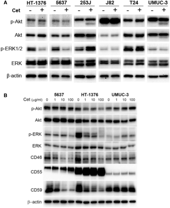
Do et al. evaluated cetuximab-mediated EGFR signaling responses in multiple bladder cancer cell lines. In Figure 2, immunoblot analysis showed that cetuximab reduced phosphorylated AKT and ERK1/2 in responsive HT1376 and 5637 cells, while several other tested lines did not show the same phosphorylation response. The data illustrate why pathway evaluation should measure defined signaling nodes in the relevant cellular context rather than infer signaling activity from antibody binding alone.
The study also demonstrates how antibody concentration, cell background, phosphoprotein readouts, and downstream biological response can be integrated to distinguish pathway-responsive from pathway-nonresponsive models. For veterinary antibody programs, Creative Biolabs can adapt this experimental logic to canine- or feline-relevant systems using phosphorylation profiling, Western blotting, phospho-sensitive cellular assays, reporter measurements, imaging, and complementary functional outputs selected around the intended target mechanism.
Advantages of Mechanism-Matched Cell Signaling Evaluation
The study is structured around what each assay can actually establish, with species context and orthogonal controls built into the experimental plan.
Species-Aware Models
Assays are adapted to canine or feline targets, cells, and available reagents.
Mechanism-Matched Readouts
Readouts are selected around the expected receptor and downstream signaling biology.
Orthogonal Evidence
Phosphorylation, localization, reporter, and output assays can support cross-checking.
Decision-Focused Reporting
Reports separate observed pathway modulation from broader efficacy or mechanism claims.
Veterinary Antibody Cell Signaling Evaluation FAQs
References
- Do, Manh-Hung, et al. "CD46 protects the bladder cancer cells from cetuximab-mediated cytotoxicity." Scientific Reports 12 (2022): 22420. https://doi.org/10.1038/s41598-022-27107-9
- Distributed under Open Access license CC BY 4.0, without modification.