
Measure Cellular Uptake Beyond Surface Binding
Antibody internalization is a distinct functional property from target binding. A candidate may bind strongly at the cell surface yet enter cells slowly, remain membrane-associated, recycle back to the surface, or traffic into intracellular compartments with different consequences for its intended mechanism. Creative Biolabs develops fit-for-purpose in vitro internalization assays that separate surface-associated signal from intracellular uptake and compare candidates under controlled conditions.
Study design can integrate kinetic flow cytometry, pH-responsive labeling, fluorescence imaging, and trafficking markers according to the target, antibody format, cell model, and research question. Readouts are interpreted in the context of antigen expression, concentration, incubation time, temperature controls, and assay background so internalization data can support candidate ranking without being mistaken for binding affinity or downstream efficacy.
Mechanism-Matched Antibody Uptake and Trafficking Evaluation
Internalization is not a single readout. We select complementary assays according to whether the project needs quantitative uptake, kinetic comparison, intracellular localization, or evidence of delivery into acidic compartments.
Quantitative Flow Cytometry Internalization Assay
Measure loss of surface-accessible antibody or gain of intracellular/internalization-associated fluorescence across defined time points. Temperature-shift, extracellular quenching, acid-strip, or differential staining strategies can be incorporated where appropriate to distinguish internalized signal from residual surface binding.
pH-Sensitive Antibody Uptake Evaluation
Use pH-responsive probes to generate increased fluorescence after antibody-associated material reaches acidic endosomal or lysosomal environments. This format is particularly useful for screening internalization and acidic-compartment trafficking relevant to ADC development. The readout is interpreted as acidification-associated uptake; it does not by itself establish efficient payload delivery, which also depends on downstream factors such as lysosomal processing, linker cleavage, antibody degradation, payload release, and payload permeability.
Imaging-Based Internalization and Trafficking Analysis
Visualize antibody redistribution from the plasma membrane into intracellular puncta using confocal or high-content imaging. When the mechanism requires localization evidence, colocalization with markers such as early endosome or lysosome-associated proteins can be incorporated to characterize trafficking patterns.
Comparative Internalization for Lead and Format Selection
Compare clones, engineered variants, isotypes, valencies, unconjugated antibodies, or ADC-relevant constructs under matched experimental conditions. Candidate ranking can integrate uptake kinetics with cell-surface target expression and binding controls to avoid attributing higher signal simply to higher antigen density or starting occupancy.
Four-Stage Veterinary Antibody Internalization Evaluation Workflow
The workflow follows the original service logic while incorporating mechanism-aware controls and readouts so uptake and trafficking data remain interpretable across candidates.
Project Consultation & Target Profiling
Define the target species, antibody format, intended mechanism, available material, target-expression context, and the candidate-selection question the study must answer.
Cell Line Selection & Assay Design
Select target-positive and relevant control cells, confirm surface expression and baseline binding, then choose flow, pH-sensitive, imaging, or combined assay formats with appropriate controls.
Internalization Assay Execution
Measure antibody uptake over defined time points and, where required, characterize intracellular trafficking using quantitative flow cytometry, confocal microscopy, or high-content imaging.
Data Analysis & Reporting
Compare internalization kinetics, localization patterns, control performance, and candidate differences, then deliver raw data, visual outputs, limitations, and method-aware interpretation.
Recommended Starting Information
- Target and intended canine or feline application
- Antibody sequence, format, concentration, and available quantity
- Target-positive cell model or preferred biological system
- Expected internalization-dependent mechanism, if known
- Comparator antibody, isotype, or reference construct availability
Typical Deliverables
- Study design and assay conditions
- Raw and processed flow or imaging data
- Time-course and concentration-response visualizations where applicable
- Comparative internalization metrics and control performance
- Method-aware interpretation and candidate ranking rationale
Choose the Readout That Matches the Internalization Question
No single assay captures every aspect of uptake. Combining a quantitative method with localization evidence is often useful when intracellular trafficking is central to candidate selection.
| Assay Approach | Primary Readout | Best Suited For | Important Interpretation Point |
|---|---|---|---|
| Surface-loss / differential-label flow cytometry | Relative surface signal or calculated internalized fraction over time | Quantitative candidate comparison and kinetic profiling | Requires appropriate controls to distinguish receptor loss, shedding, or assay artifacts from true uptake. |
| pH-sensitive fluorescence | Fluorescence increase in acidic intracellular compartments | Screening internalization and acidic-compartment trafficking relevant to ADC development | Reports acidification-associated trafficking and may not capture antibody residing in non-acidic compartments. |
| Confocal / high-content imaging | Spatial redistribution and intracellular puncta | Visual confirmation, trafficking pattern, and organelle colocalization | Image analysis should use matched acquisition settings and objective segmentation rather than representative images alone. |
| Orthogonal combined workflow | Quantitative uptake plus localization evidence | Mechanism-focused lead selection and format comparison | Interpretation should account for target density, starting binding, concentration, and cell-model differences. |
Need to compare internalization across several antibody leads?
Share your target, antibody format, available cell system, and intended mechanism. We can recommend a focused screening design and the controls needed for interpretable candidate ranking.
Published Data on Antibody Internalization and Lysosomal Trafficking
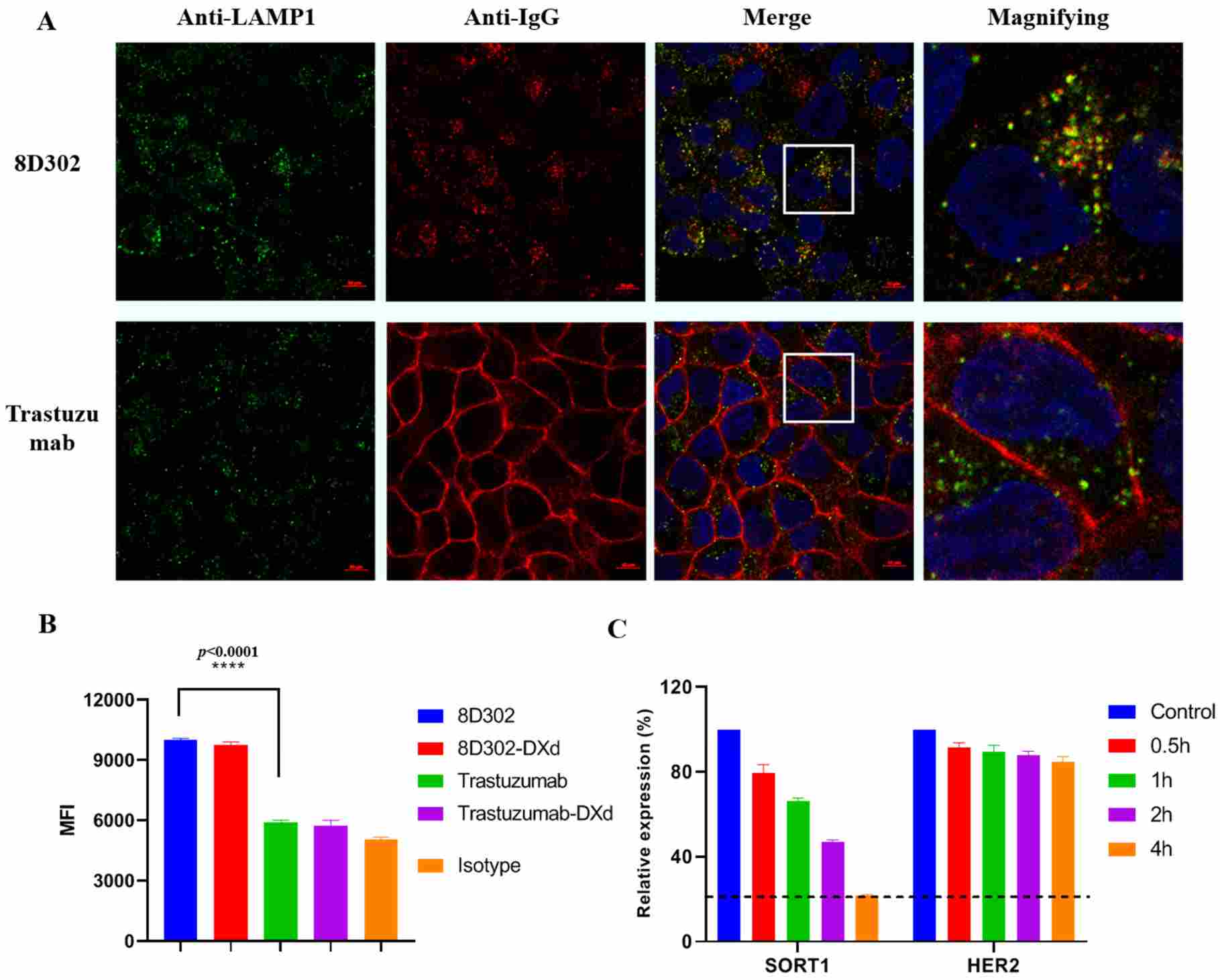
Zhuang et al. compared the internalization behavior of a SORT1-targeting antibody with trastuzumab in T47D cells. Their Figure 7 combined confocal microscopy with pH-sensitive flow cytometry: microscopy visualized intracellular antibody and lysosomal colocalization, while pHrodo-based flow cytometry provided a quantitative readout associated with trafficking into acidic endosomal or lysosomal compartments.1
The study illustrates why internalization evaluation benefits from complementary readouts rather than surface binding alone. Creative Biolabs can apply the same experimental logic to veterinary antibody programs by combining target-expression controls, quantitative uptake assays, pH-sensitive formats, and imaging-based localization to compare canine or feline candidates and support ADC-oriented or receptor-modulating mechanism studies.
Why Choose Creative Biolabs for Antibody Internalization Evaluation?
Study design is adapted to the target, antibody format, and intended mechanism, with emphasis on controls and readouts that support defensible comparison.
Veterinary-Relevant Models
Canine and feline cell systems can be incorporated when suitable reagents and models are available.
Orthogonal Readouts
Flow and imaging approaches can separate quantitative uptake from intracellular localization evidence.
Mechanism-Aware Controls
Target expression, temperature, background, and binding controls strengthen internalization interpretation.
Decision-Oriented Reporting
Comparative datasets are organized to support lead ranking and downstream assay planning.
Veterinary Antibody Internalization Evaluation FAQs
References
- Zhuang, Weiliang, et al. "Generation and Characterization of SORT1-Targeted Antibody–Drug Conjugate for the Treatment of SORT1-Positive Breast Tumor." International Journal of Molecular Sciences 24.24 (2023): 17631. https://doi.org/10.3390/ijms242417631
- Distributed under Open Access license CC BY 4.0, without modification.