
Functional Evidence Beyond Antibody Binding
Target binding alone does not establish whether a veterinary therapeutic antibody produces the intended biological effect. Creative Biolabs develops fit-for-purpose in vitro assays that connect target engagement with mechanism-specific outcomes such as pathway blockade, receptor internalization, signaling modulation, altered cell fate, or Fc-mediated effector activity.
Each study is configured around the target species, antibody format, mechanism of action, available cell systems, and the decision the data must support. Dose-response design, matched controls, orthogonal readouts, and transparent interpretation help distinguish functional potency from simple binding and create a stronger basis for lead ranking and downstream preclinical planning.
Mechanism-Matched In Vitro Functional Assay Services
We select functional endpoints according to the antibody's intended mechanism rather than applying one generic potency assay. Individual modules can be used for screening, lead comparison, mechanism confirmation, or assay-development support.
Blocking and Neutralization Evaluation
Assess whether a candidate disrupts a ligand-receptor, receptor-cofactor, pathogen-host, or other defined biological interaction. Cell-free competition can establish blocking behavior, while cell-based assays test whether that inhibition produces the expected downstream response. Neutralization studies can be adapted to soluble mediators or pathogen-related systems when an appropriate model is available.
Antibody Internalization Evaluation
Measure the time-dependent uptake of target-bound antibody using imaging, flow cytometry, or other quantitative internalization approaches. Assay design can compare temperature, time, concentration, target density, or candidate format and can distinguish surface-associated signal from internalized material. For payload-delivery programs, internalization is interpreted as one component of the delivery mechanism rather than proof of intracellular payload release.
Cell Signaling Pathway Evaluation
Determine whether antibody treatment activates, suppresses, or redirects a defined signaling pathway. Depending on target biology, studies may use phospho-protein analysis, Western blotting, phospho-flow, transcriptional reporters, or secreted biomarkers. Time-course and concentration-response designs help separate early receptor-proximal signaling from later secondary cellular effects.
Cell-Based Functional Response Evaluation
Quantify direct effects on cell behavior when proliferation, growth inhibition, apoptosis, viability, activation, differentiation, or cytokine production is part of the intended mechanism. Relevant target-positive and control cells are incorporated wherever possible so that candidate activity can be interpreted in the context of target dependence and assay background.
Fc Effector Function Evaluation
Characterize Fc-dependent activity using ADCC, CDC, ADCP, or related formats selected for the intended veterinary antibody mechanism. Species-relevant Fc receptor, complement, effector-cell, and target-cell context is considered when suitable reagents or models are available. Fc-silent, isotype, target-negative, or other mechanism controls can be incorporated to clarify whether the observed response is truly Fc dependent.
Match the Functional Question to the Right Readout and Controls
Functional potency is meaningful only when the assay model, readout, and controls reflect the proposed mechanism. The matrix below shows representative options used to build a decision-oriented assay plan.
| Functional Question | Representative Assay Approaches | Important Controls | Decision-Oriented Output |
|---|---|---|---|
| Does the antibody block a biological interaction? | Competition ELISA, receptor-ligand inhibition, reporter cells, cytokine or pathway readout. | No-antibody, isotype, ligand-only, positive inhibitor, target-negative system where feasible. | Percent inhibition, IC50, maximal inhibition, candidate rank order. |
| Does the antibody internalize after target binding? | Flow cytometry, high-content imaging, pH-sensitive probes, time-course uptake analysis. | 4 C surface-binding control, target-negative cells, unconjugated or nonbinding control. | Internalized fraction, uptake kinetics, concentration and target-density dependence. |
| Does target engagement alter signaling? | Phospho-flow, Western blot, reporter gene assays, transcriptional or secreted biomarker analysis. | Baseline, pathway agonist or inhibitor, isotype, target-negative or knockdown control when appropriate. | Direction and magnitude of pathway modulation, EC50/IC50, time dependence. |
| Does the antibody change cell fate or behavior? | Apoptosis, proliferation, growth inhibition, viability, activation, cytokine release. | Vehicle/no-antibody, isotype, positive control, target-negative comparator. | Functional potency, maximal effect, target dependence, comparative lead matrix. |
| Is activity mediated through the Fc region? | ADCC, CDC, ADCP, Fc-receptor reporter assays or primary-effector formats. | Fc-silent or isotype control, effector-only, target-only, antigen-negative target cells. | Fc-dependent activity profile, potency, maximal response, format comparison. |
From Mechanism Definition to Decision-Ready Functional Data
Our stage-gated workflow keeps assay design, species context, controls, execution, analysis, and reporting aligned with the biological question being tested, so each study phase produces evidence that can guide the next project decision.
Recommended Starting Information
- Target name, sequence, and intended species
- Antibody format and Fc design
- Expected mechanism of action
- Available target-positive and control cells
- Existing binding or specificity data
- Preferred decision criteria and downstream study
Typical Final Deliverables
- Customized assay plan and acceptance criteria
- Raw and processed functional datasets
- Dose-response and potency analysis
- Control and target-dependence interpretation
- Candidate comparison and rank-order summary
- Methods, results, conclusions, and next-step recommendations
Need a Functional Assay Strategy for a Veterinary Antibody Candidate?
Share the target species, antibody format, proposed mechanism, and current binding data. We can help map the most informative functional endpoints and controls.
Published Data: Canine PD-1/PD-L1 Blockade Restores a Functional T-Cell Readout
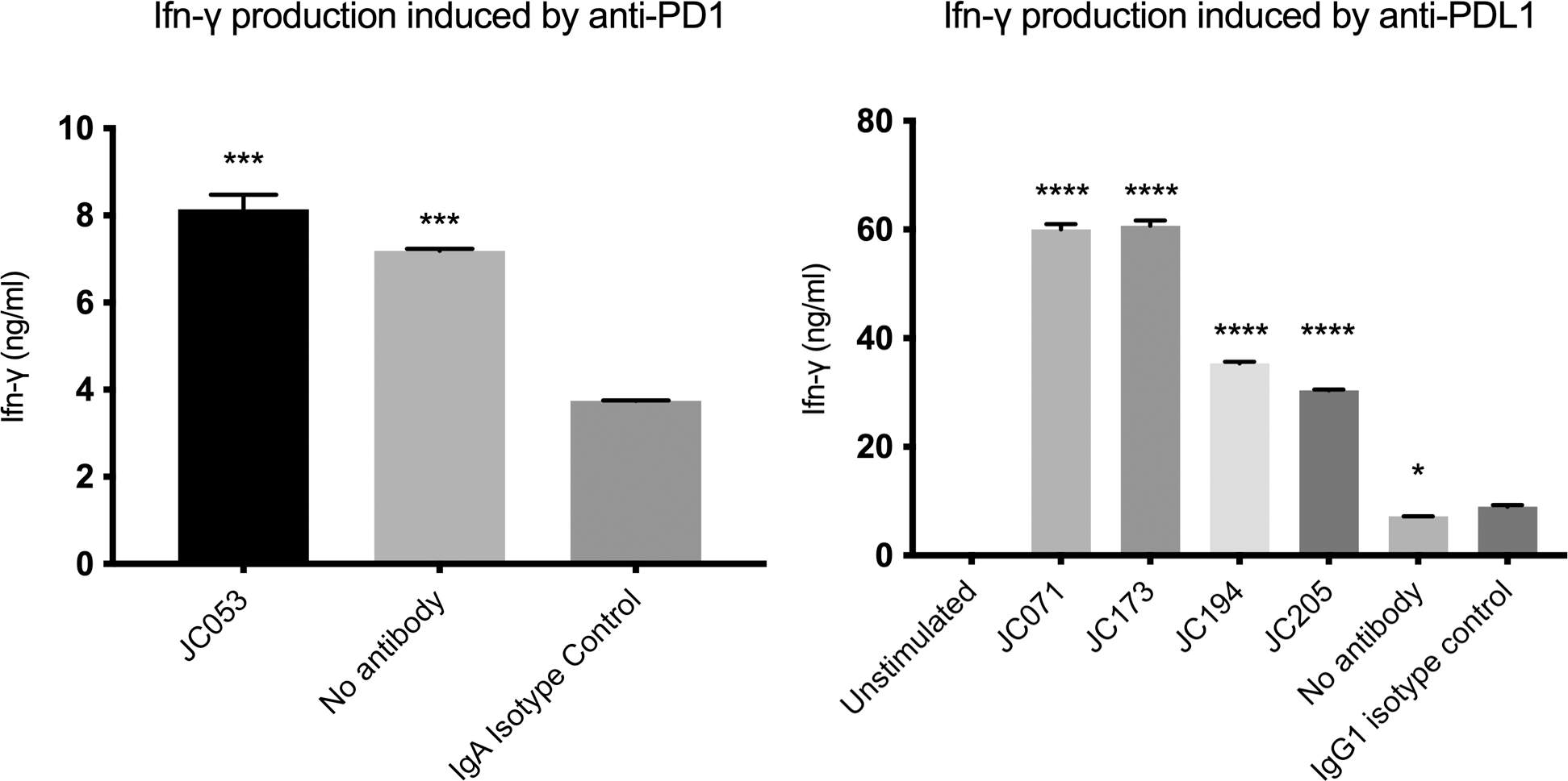
Choi et al. evaluated canine PD-1/PD-L1 monoclonal antibodies using a canine peripheral blood mononuclear cell assay. In Figure 8 of the source article, ConA-stimulated PBMC cultures treated with several PD-L1-blocking antibodies produced significantly more IFN-γ than the corresponding IgG1 isotype control, demonstrating that a candidate's biological activity can be distinguished from binding alone by measuring a downstream immune-cell response.
This study illustrates a useful functionality-testing principle for veterinary antibody programs: pair target or ligand blocking evidence with a species-relevant cell system and a quantitative downstream readout. Creative Biolabs can apply the same mechanism-first logic to blocking, signaling, cytokine, cell-fate, internalization, or Fc-effector assays, with dose-response analysis and matched controls selected for the specific candidate and decision point.
Why Choose Creative Biolabs for Veterinary Antibody Functional Evaluation?
Our assay-development approach emphasizes species context, mechanism alignment, quantitative controls, and interpretation that remains within the evidence supported by the selected model.
Species-Aware Assay Design
Match cell models, reagents, and controls to canine or feline biology.
Mechanism-First Strategy
Select functional endpoints that directly test the intended antibody mechanism.
Flexible Readout Platforms
Combine flow cytometry, imaging, reporter, cytokine, and cytotoxicity measurements.
Decision-Ready Reporting
Compare candidates with transparent controls, potency metrics, and interpretation boundaries.
Frequently Asked Questions About In Vitro Biological Functionality Evaluation
Binding evaluation asks whether an antibody recognizes its target and can quantify binding behavior with an appropriate method. Biological functionality evaluation asks what happens after target engagement, such as pathway blockade, signaling change, internalization, altered cell fate, cytokine modulation, or Fc-mediated effector activity. The two evidence types are complementary but should not be treated as interchangeable.
A project can combine blocking or neutralization, internalization, signaling, cell-fate, cytokine, ADCC, CDC, ADCP, and related assays when those endpoints fit the proposed mechanism. We generally prioritize a focused assay cascade rather than running every platform, so each experiment contributes to a defined candidate decision.
Yes. Client-supplied antibodies, target cells, engineered lines, antigens, ligands, effector cells, or reference reagents can be reviewed for suitability and incorporated when appropriate. The study plan should document material identity, handling requirements, assay compatibility, and the controls needed for interpretable comparisons.
The assay format depends on the antibody Fc, target antigen, species context, and available reagents. ADCC, CDC, ADCP, or Fc-receptor reporter approaches may be considered, with target-positive and negative cells plus Fc-silent, isotype, effector-only, or other controls used where appropriate to establish Fc dependence.
No. EC50 and IC50 values from cell-based or pathway assays are assay-dependent measures of functional potency. Equilibrium affinity and kinetic parameters such as KD, kon, and koff require an appropriate binding method, typically SPR or BLI when experimentally suitable. Functional potency can be compared with binding data, but the metrics should remain distinct.
Useful starting information includes the veterinary species, target, antibody sequence or format, Fc design, proposed mechanism of action, existing binding or specificity data, available cell models or reagents, and the decision the assay must support. These inputs help determine whether a pilot assay, focused potency study, or broader functional panel is most appropriate.
References
- Choi, Jin Wook, et al. "Development of canine PD-1/PD-L1 specific monoclonal antibodies and amplification of canine T cell function." PLOS ONE 15.7 (2020): e0235518. https://doi.org/10.1371/journal.pone.0235518
- Distributed under Open Access license CC BY 4.0, without modification.