
Confirm Veterinary Antibody Target Engagement in a Cellular Context
In vitro cell binding evaluation asks a practical early-development question: does the antibody recognize its intended target when that target is presented by a relevant cell? Creative Biolabs designs canine- and feline-focused binding studies around target expression, membrane topology, antibody format, available reagents, and the decision the data must support.
Assay plans center on flow cytometry for single-cell binding profiles and cell-based ELISA for scalable comparative screening, with immunoprecipitation available as a complementary method for orthogonal target-capture confirmation. Results are interpreted as cellular target-engagement evidence and candidate-ranking data. When true kinetic affinity parameters are required, complementary SPR or BLI studies can be incorporated rather than treating ELISA or routine flow-cytometric readouts as direct measurements of kon and koff.
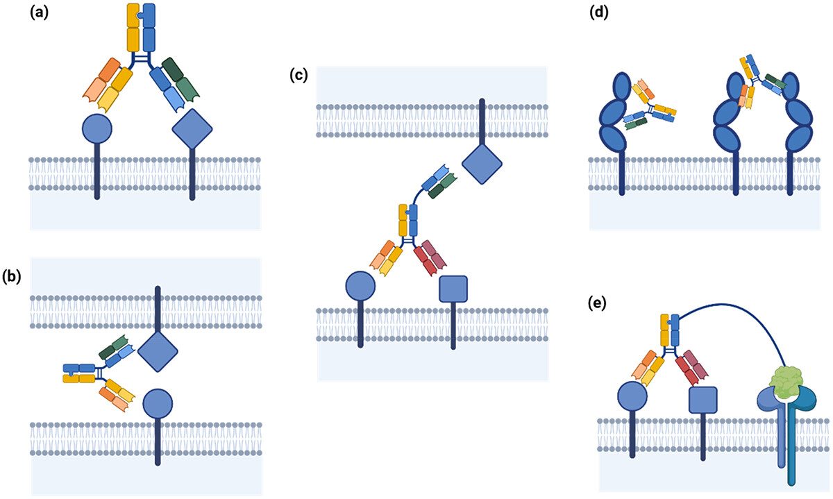
Flow Cytometry for Quantitative Cell Binding Profiles
Measure binding across individual cells while controlling for antigen expression, background signal, and population heterogeneity.
Target-Positive Cell Binding
Titrated antibody binding to naturally expressing or engineered canine and feline cells, with matched negative controls where appropriate.
→Candidate Ranking and Apparent Binding Activity
Concentration-response curves using MFI, percent-positive cells, background-corrected signal, and EC50-style comparative metrics.
Competition and Cross-Species Comparisons
Optional competition, ortholog, or cell-background comparisons help clarify relative target engagement before dedicated specificity studies.
→Cell-based ELISA for Comparative Binding Assessment
Use a plate-based whole-cell format when throughput and consistent population-level comparison are more important than single-cell resolution.
Whole-Cell Binding Screens
Evaluate antibody binding across candidate panels using target-expressing cells, optimized detection reagents, and appropriate background controls.
→Dose-Response and Signal Window Optimization
Optimize cell density, antibody concentration, incubation conditions, detection chemistry, and signal-to-background performance before comparative testing.
→Immunoprecipitation and Target-Capture Support
Complement cell-surface binding with biochemical evidence that the antibody captures the expected cell-derived target or target complex.
Immunoprecipitation from Cell Lysates
Evaluate target capture under defined lysis and wash conditions, followed by western blot or other compatible detection approaches.
→Orthogonal Identity Checks
Use target-positive and negative materials, isotype controls, input controls, and expected molecular size to strengthen interpretation.
Veterinary Antibody In Vitro Cell Binding Evaluation Workflow
A six-stage workflow connects cell-model quality, assay controls, concentration-response design, and orthogonal confirmation so each result can be interpreted in the context of the project decision.
Recommended Starting Information
- Target name, sequence, and species context
- Antibody sequence, format, concentration, and purity
- Available target-positive and negative cells
- Known expression or prior binding data
- Desired ranking or go/no-go criteria
- Planned downstream functional studies
Typical Final Deliverables
- Assay design, controls, and acceptance criteria
- Raw and processed cytometry or plate data
- Binding curves and comparative metrics
- Gating strategy or plate-quality summaries
- Orthogonal target-capture evidence when included
- Interpretation and next-step recommendations
Match the Cell Binding Question to the Right Evaluation Method
No single assay answers every binding question. Selecting the method by readout and limitation prevents apparent binding activity, biochemical target capture, and true kinetic affinity from being treated as interchangeable measurements.
| Method | Best Used For | Representative Readouts | Interpretation / Limitation |
|---|---|---|---|
| Flow Cytometry | Cell-surface target engagement and candidate ranking at single-cell resolution. | MFI, percent-positive cells, signal-to-background, binding curve, apparent EC50. | Excellent for heterogeneous populations and expression gating; routine assays do not directly provide kon/koff. |
| Cell-based ELISA | Plate-based comparative screening across larger candidate or condition sets. | OD or luminescent signal, background-corrected response, apparent EC50, rank order. | Population-average readout; limited single-cell information and not a direct kinetic measurement. |
| Immunoprecipitation | Orthogonal capture of native or cell-derived target from lysate. | Target enrichment, expected band or detection signal, control comparison. | Supports target identity/capture; outcome depends on epitope accessibility, lysis conditions, and complex stability. |
| SPR / BLI (Complementary) | Quantitative real-time interaction analysis using a suitable purified binding system. | KD, kon, koff where experimentally appropriate. | Provides kinetic affinity information but may not reproduce cell-surface density, avidity, or membrane context. |
Start with the decision your binding data must support
Creative Biolabs can recommend a focused single-assay study or an orthogonal binding package after reviewing your target, species, cells, antibody format, and downstream development plan.
Published Data on Canine CD20 Antibody Binding Validation
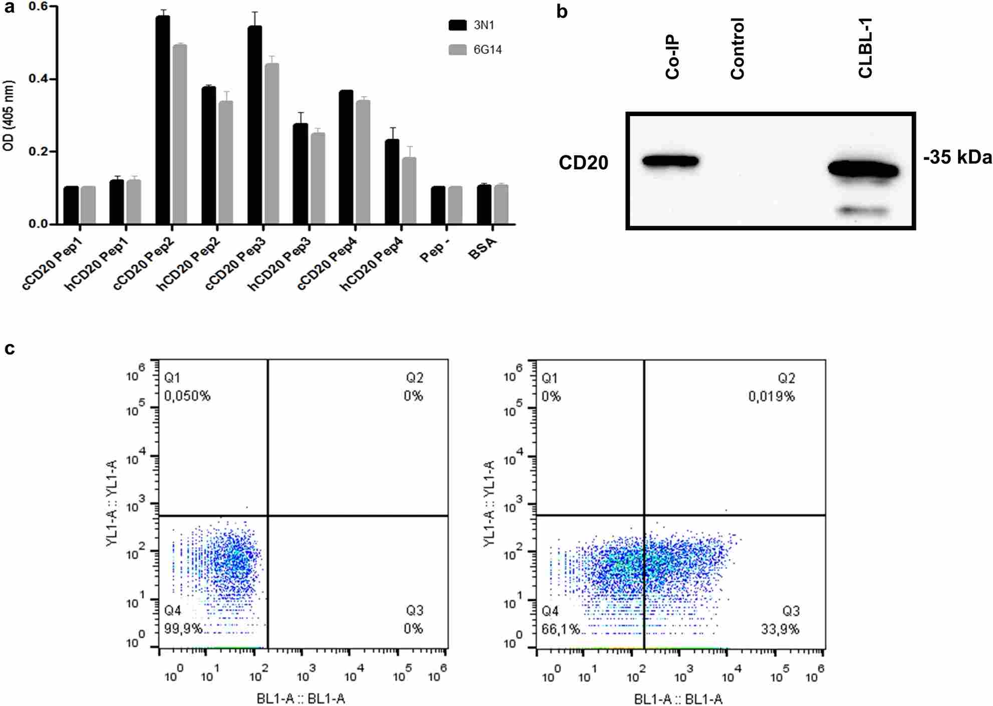
Dias et al. characterized rabbit-derived single-domain antibodies against canine CD20 using complementary binding approaches. The reported figure combines peptide ELISA, co-immunoprecipitation of CD20 from canine lymphoma cell extracts, and live-cell flow cytometry, illustrating how orthogonal methods can connect epitope recognition, biochemical target capture, and binding to a native cell-surface target in a veterinary setting.
Although the ELISA component in this published example was used for peptide-based epitope mapping rather than whole-cell binding, the study illustrates the value of combining orthogonal binding and target-confirmation methods. Creative Biolabs can configure flow-cytometric cell binding and cell-based ELISA as core cellular binding assays, with immunoprecipitation used where appropriate for orthogonal target confirmation and SPR or BLI added when direct affinity or kinetic measurements are required.
Why Choose Creative Biolabs for Veterinary Antibody Cell Binding Evaluation
Assay design stays anchored to species context, cell biology, controls, and the specific candidate decision your project needs to make.
Species-Relevant Models
Canine and feline cell systems are selected around target expression and project biology.
Method-Aware Interpretation
Cell binding, biochemical capture, and kinetic affinity are reported as distinct evidence types.
Orthogonal Assay Options
Flow cytometry, cell ELISA, IP, and biophysical methods can be combined strategically.
Decision-Ready Reporting
Controls, raw data, analysis, limitations, and candidate-ranking conclusions remain traceable.
Build a Cell Binding Study Around Your Veterinary Antibody Decision Point
Whether you need a focused flow-cytometry comparison, a plate-based screen, orthogonal immunoprecipitation evidence, or a broader binding package, our team can align the study with your target biology, available material, species context, and downstream evaluation plan.
Veterinary Antibody In Vitro Cell Binding Evaluation FAQs
References
- Lodge, James, et al. "Quantifying antibody binding: techniques and therapeutic implications." mAbs 17.1 (2025): 2459795. https://doi.org/10.1080/19420862.2025.2459795
- Dias, Joana N. R., et al. "Characterization of the canine CD20 as a therapeutic target for comparative passive immunotherapy." Scientific Reports 12 (2022): 2678. https://doi.org/10.1038/s41598-022-06549-1
- Distributed under Open Access license CC BY 4.0, without modification.