
In Vitro Specificity & Cross-Reactivity Evaluation Service for Veterinary (Pet) Antibody

Creative Biolabs designs species-aware in vitro specificity and cross-reactivity studies for canine and feline therapeutic antibodies, integrating cell-based binding, related-target counter-screening, ortholog comparison, tissue cross-reactivity, and orthogonal confirmation to support informed lead selection and preclinical risk assessment.

Service Overview

Define Antibody Selectivity Before Off-Target Signals Become a Development Liability

Specificity and cross-reactivity are not a single readout. A veterinary antibody may bind its intended antigen strongly yet also recognize a related family member, a species ortholog, a conformationally similar surface protein, or an unexpected tissue-associated epitope. Creative Biolabs builds in vitro evaluation programs around these distinct questions rather than relying on one screening format.

Study design can combine target-positive and target-negative cells, related-protein or ortholog panels, concentration-dependent binding, species-relevant tissues, and orthogonal confirmation. The resulting evidence helps distinguish target-associated binding from assay background, broad family reactivity, and potentially biologically relevant off-target binding. Early identification of cross-reactivity can help deprioritize problematic candidates before additional functional, PK/PD, or in vivo resources are committed.

Evaluation Scope

Species-Relevant Specificity and Cross-Reactivity Testing for Veterinary Antibody Candidates

The assay cascade is configured around target biology, epitope context, intended species, antibody format, available reagents, and the decision that must be made. Modules can be used independently or combined into a staged specificity package.

Target-Positive and Target-Negative Cell Specificity

Compare binding on cells that differ in target expression to establish a practical selectivity baseline in a native or engineered cellular context. Because absent signal in a target-negative system does not by itself prove molecular specificity, the control strategy can be strengthened where appropriate to distinguish target-dependent binding from Fc receptor effects, detection background, transfection artifacts, or nonspecific membrane association.

  • Flow cytometry or cell-based plate formats
  • Target-negative, matched mock-transfected, or target-knockout controls where available
  • Fc-receptor blocking, detection-only, or isotype controls where relevant
  • Competition or blocking studies for ambiguous binding
  • Concentration-response comparison and background assessment
Related-Protein and Family-Member Counter-Screening

Test structurally or functionally related antigens that could create unintended pharmacology or complicate interpretation of downstream functional studies.

  • Paralogs and homologous target families
  • Recombinant protein or cell-displayed formats
  • Competitive or confirmatory formats where appropriate

Cross-Species and Ortholog Reactivity Assessment

Determine whether a candidate recognizes orthologous targets from the intended veterinary species and selected comparison species without assuming sequence identity predicts binding.

  • Canine and feline ortholog comparison
  • Additional species selected by program need
  • Species-specific detection reagents and controls

Tissue Cross-Reactivity and Off-Target Binding

Extend screening into normal species-relevant tissue sections or tissue microarrays when cellular or protein panels cannot reproduce native architecture and epitope presentation.

  • Multi-organ canine or feline tissue panels
  • Immunohistochemical signal localization
  • Pattern review with matched controls and concentration context

Orthogonal Specificity Confirmation

Resolve ambiguous signals by using a method with a different detection principle, antigen format, or cellular context rather than repeating the same assay.

  • Cell-based and protein-based cross-checking
  • Competition or blocking experiments
  • Target-capture or biochemical confirmation when scientifically appropriate

Integrated Candidate Comparison and Interpretation

Bring cell, ortholog, related-target, and tissue findings into one decision framework that highlights reproducible signals and remaining uncertainties.

  • Candidate-by-candidate selectivity profiles
  • Flagged off-target observations and follow-up priorities
  • Methods, raw and analyzed data, and interpretation summary
Workflow

A Five-Stage Specificity and Cross-Reactivity Evaluation Workflow

A stage-gated workflow keeps target context, panel selection, assay controls, quantitative comparison, and follow-up interpretation traceable from study design through reporting.

01
Project Review & Epitope ContextDefine species, target, mechanism, epitope information, related proteins, and orthologs.
02
Panel Design or SourcingSelect or develop target-positive, negative, homologous, orthologous, and tissue test systems.
03
Assay Development & ControlsOptimize signal range, antibody concentrations, detection reagents, and fit-for-purpose controls such as target-negative, mock-transfected, knockout, detection-only, Fc-blocking, or isotype controls where appropriate.
04
Comparative Binding AnalysisQuantify target selectivity, off-target signals, and interspecies reactivity across the defined panel.
05
Reporting & Follow-UpIntegrate curves, panel results, flagged risks, interpretation, and recommended confirmation studies.
Assay Matrix

Match the Cross-Reactivity Question to the Appropriate Test System

Different off-target questions require different biological contexts. A protein panel can be efficient for family-member screening, whereas cells or tissue sections may be more informative when conformation, membrane organization, or native tissue architecture matters.

Evaluation Question Representative Test System Key Controls / Readouts Decision-Oriented Output
Intended Target Specificity Target-positive versus target-negative, matched mock-transfected, or target-knockout cells where available Concentration series; target-negative or mock/knockout controls; detection-only, Fc-blocking, or isotype controls where relevant; signal intensity and positive-cell fraction Evidence that binding tracks with target expression, with control data to help qualify assay background and other non-target-mediated signals
Related-Target Selectivity Protein family panel or cells expressing selected paralogs Matched antigen loading or expression, comparative dose-response, competition where useful Identification of family-member binding and a prioritized confirmation plan
Species / Ortholog Reactivity Canine, feline, and selected ortholog proteins or cells Species-matched reagents, comparable expression, relative binding across orthologs Cross-species reactivity profile for model and development planning
Tissue Off-Target Binding Normal canine or feline tissue sections or tissue microarrays Positive and negative controls, concentration context, cellular localization, staining pattern Map of unexpected tissue-associated binding for follow-up assessment
Ambiguous or Borderline Signal Orthogonal cell, protein, competition, or biochemical method Independent detection principle and prespecified acceptance logic Confirmation, rejection, or qualification of the original observation

Start with your antibody, target, species, and current binding data

Our scientists can define a focused counter-screen or a broader cell, ortholog, and tissue cross-reactivity package based on the evidence needed for your next candidate decision.

Peer-Reviewed Veterinary Evidence

Published Data on Antibody Cross-Reactivity Testing in Feline Leukocytes

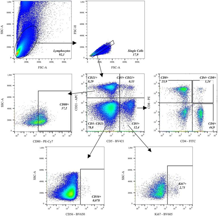
Krüger et al. evaluated 72 monoclonal antibodies for reactivity with feline leukocytes and reported cross-reactivity for 35 antibodies spanning CD markers, a chemokine receptor, cytokines, and other targets. The study used flow cytometry to distinguish labeled feline lymphocyte populations, and the illustrated gating strategy shows how marker combinations and controlled population definition can support interpretable species-specific antibody testing.

Although the study focused on research antibodies rather than therapeutic leads, its experimental logic is directly relevant to veterinary specificity assessment: cross-reactivity should be demonstrated empirically in the intended species, interpreted with appropriate controls, and confirmed in a biologically meaningful cellular context. Creative Biolabs applies the same principles when designing target-positive/negative comparisons, ortholog panels, related-protein counter-screens, and follow-up cell or tissue assays for canine and feline therapeutic antibody candidates.

Flow cytometry gating of feline lymphocyte subsets with species-relevant antibody markers. (OA Literature)
Fig.1 Flow-cytometric gating strategy for feline lymphocyte populations using validated antibody markers.1,2
Service Advantages

Why Choose Creative Biolabs for Veterinary Antibody Specificity Evaluation?

Species relevance, assay context, and orthogonal evidence are built into the study design so cross-reactivity findings are useful for candidate comparison rather than isolated screening results.

Veterinary antibody specificity and cross-reactivity evaluation at Creative Biolabs

Veterinary-Specific Focus

Canine and feline systems keep selectivity decisions aligned with intended species biology.

Customized Evaluation Strategy

Panel design follows target family, epitope context, mechanism, and downstream study needs.

Layered Risk Assessment

Cell, protein, ortholog, and tissue evidence reduces single-assay interpretation risk.

Decision-Oriented Reporting

Integrated datasets highlight off-target signals, species differences, and next-step priorities.

Frequently Asked Questions

Veterinary Antibody Specificity & Cross-Reactivity FAQs

Canine and feline systems are a central focus. Additional veterinary or comparison species can be discussed when suitable antigens, cell systems, tissues, and detection reagents are available. Species selection should follow the intended therapeutic species, target conservation, model strategy, and the specific cross-reactivity question.

References

  1. Krüger, H., et al. "Flow cytometric evaluation of monoclonal antibodies for cross-reactivity with feline leukocytes." Frontiers in Veterinary Science 13 (2026): 1778256. https://doi.org/10.3389/fvets.2026.1778256.
  2. Distributed under Open Access license CC BY 4.0, without modification.

This site is protected by reCAPTCHA and the Google Privacy Policy and Terms of Service apply.