Creative Biolabs is offering the most comprehensive services for antibody development projects. With strict regulation and effective execution, we are dedicated to providing the most valuable solutions to complete your projects.
HighlightsWith over a decade of experience in phage display technology, Creative Biolabs can provide a series of antibody or peptide libraries that are available for licensing or direct screening. These ready-to-use libraries are invaluable resources for isolating target-specific binders for various research, diagnostic or therapeutic applications. Highlights
Creative Biolabs has established a broad range of platforms for developing novel antibodies or equivalents. These cutting-edge technologies enable our scientists to meet your demands from different aspects and tailor the most appropriate solution that contributes to the success of your projects.
HighlightsWith deep understanding in antibody-related realms and extensive project experience, Creative Biolabs offers a variety of references to help you learn more about our capacities and achievements, including infographic, flyer, case study, peer-reviewed publications, and all kinds of knowledge that can assist your projects. You are also welcome to contact us directly for more specific solutions.
HighlightsGet a real taste of Creative Biolabs, one of the most professional custom service providers in the world. We are committed to providing highly customized comprehensive solutions with the best quality to advance your projects.
HighlightsHuman monoclonal antibodies offer unparalleled advantages, for example, the ability to effectively manipulate their effector functions while avoiding the immunogenicity of rodent antibodies, which are undoubtedly the best choice in disease treatment. However, the technology to generate human monoclonal antibodies is still not fully available. Creative Biolabs has extensive experience in producing human monoclonal antibodies. Based on our specialized technological platform and professionalism, our human monoclonal antibody production services are ethically certified and cover different processes, such as Native™ Human Antibody Discovery Service, and human Antibody Production Using Transgenic Mice.
In this study, anti-CLDN3 scFv was isolated by phage display using CHO-K1 cells expressing human CLDN3 (hCLDN3/CHO-K1) and human CLDN3-embedded lipid particles as antigens to create a monoclonal antibody (mAb) that recognizes CLDN3. The selection of scFv was monitored by measuring the ratio of output to input and ELISA assay, which showed the extent of scFv enrichment for CLDN3. Among the 190 scFv clones selected from hCLDN3/CHO-K1 cell translations, a 4G3 clone that showed highly specific binding to CLDN3 by flow cytometry was selected. In hCLDN3-embedded lipo-particle panning, the 190 clones were screened by lipo-particle-based ELISA and 165 clones were obtained, besides, sequencing results confirmed that all of them were consistent with the 4G3 clone. The 4G3 scFv clones were transformed into human IgG1 (h4G3) and purified by protein A affinity chromatography. SDS-PAGE analysis of the integrity of h4G3 detected the correct size of IgG heavy and light chains and full IgG at 50 kDa, 25 kDa, and 150 kDa, respectively. To verify recognition in cancer cells, CLDN3 expression was confirmed, and based on CLDN3 expression, cell-surface h4G3 binding was observed in various cancer cell lines (Fig. 1). Taken together, these findings verified the successful isolation of the scFv clone (4G3) and the production of a human monoclonal antibody (h4G3) that recognized the conformational structures of hCLDN3 and mCLDN3 and did not cross-react with other CLDNS.
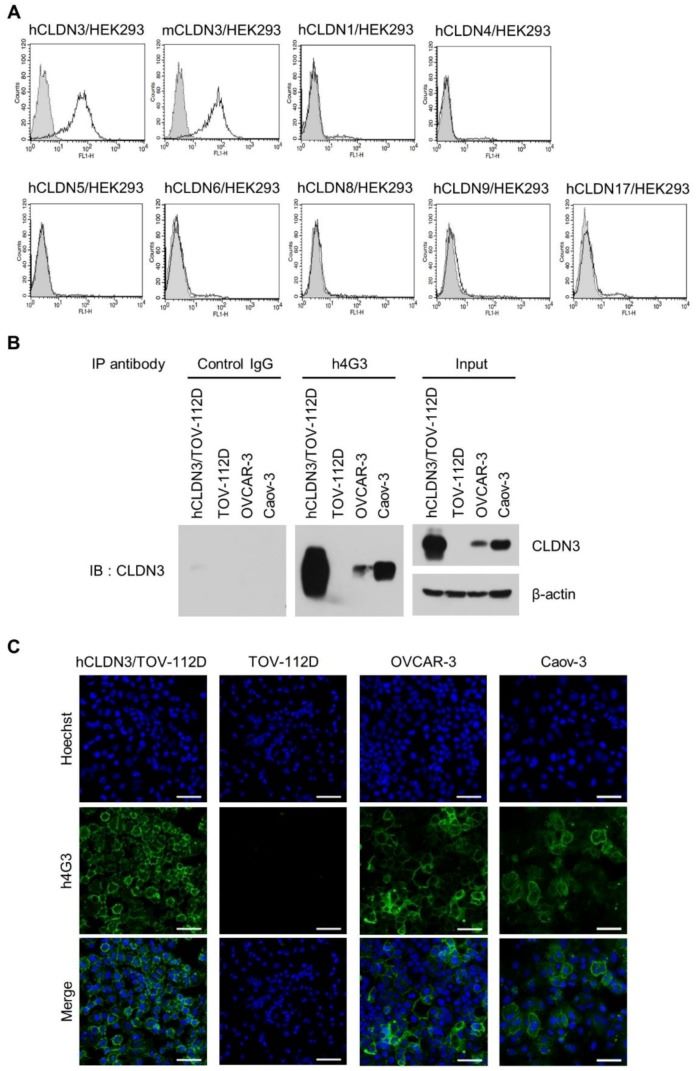 Fig 1. Specificity and conformational structure recognition of h4G3 against claudin-3 (CLDN3). (A) Stable CLDN-expressing HEK293 cells were stained with h4G3 and detected by flow cytometry. (B) The cell lysates were prepared by a probe sonicator in PBS buffer and precipitated with h4G3 or control human IgG. (C) hCLDN3/TOV-112D, TOV-112D, OVCAR-3, and Caov-3 cells were incubated with h4G3 for 1 h at 4 °C, fixed, and stained with fluorescein isothiocyanate (FITC)-conjugated goat anti-human IgG. (Yang, H., et al., 2019)
The results (Fig. 2) of the combined CPE binding study, confirmation of chimeric fusion protein expression by western blotting and other assays, suggest that h4G3 recognizes the conformational structure of the ECL2 structural domain of CLDN3 on the cell membrane and binds to MCLDN3, as there is only a 1 aa difference in ECL2 between hCLDN3 (150V) and MCLDN3 (150L).
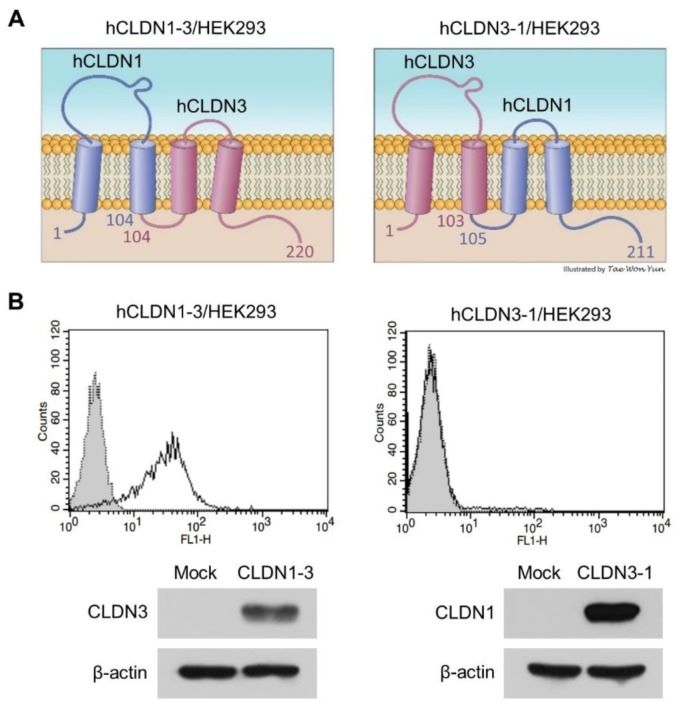 Fig 2. Binding of h4G3 to the second extracellular loop of CLDN3. (A) Structure of extracellular domain fusion CLDNs. (B) Control human IgG (gray closed dotted histogram) and h4G3 (open solid histogram) were added to hCLDN1-3/HEK293 and hCLDN3-1/HEK293 cells, and the bound antibodies were detected by flow cytometry using FITC-conjugated goat anti-human IgG. (Yang, H., et al., 2019)
The binding kinetics of h4G3 to CLDN3 on the cell surface was assessed by LigandTracer Green (Ridgeview Instruments AB) to measure real-time binding kinetics on live cells. The experimental results showed that h4G3 bound hCLDN3 on the cell membrane with typical antibody affinity and had a lower affinity for mCLDN3.
The present study evaluated the inhibitory activity of the ADCC of h4G3 on different cancer cells and showed that the cytotoxic activity of h4G3 was mediated by ADCC according to the expression of CLDN3, independent of cancer type.
In this study, subcutaneous OVCAR-3 and T47D xenograft models were established to investigate whether h4G3 could recognize CLDN3-overexpressing tumors in vivo. The distribution of h4G3 in mice was also monitored by CF750 fluorescent dye to assess the tumor targeting of h4G3. Finally, the antibody distribution in mouse organs was examined to determine the accumulation effect of h4G3 in vivo. The results showed that h4G3 is highly specific for CLDN3-expressing tumors and is therefore suitable for the detection of aberrantly expressed CLDN3 on tumor tissue, which indicates the potential value of h4G3 as a diagnostic and CLDN3-targeted therapy for cancer.
Reference
All listed services and products are For Research Use Only. Do Not use in any diagnostic or therapeutic applications.