Custom Anti-GD3 Antibody Service for Melanoma & Neuroectodermal Tumor Research
Custom Anti-GD3 Antibody Development Service
Creative Biolabs develops anti-GD3 antibodies with tailored antigen presentation,
counter-screening, and cell-based validation to improve specificity across structurally related
gangliosides and support reliable research assays.
Plan the Antibody Around How GD3 Is Really Presented
Creative Biolabs helps researchers develop anti-GD3 antibodies around the way this
disialoganglioside is actually displayed: as a compact, sialylated headgroup anchored in a lipid membrane.
Because GD3 shares biosynthetic and structural features with GD2, GM3, and modified GD3 species, a useful
program must do more than generate a strong plate signal. We work with you to align antigen presentation,
antibody format, counter-screening, and cell-based confirmation with the intended research assay.
GD3 is a demanding antibody target for a simple reason: the
epitope is not only a carbohydrate sequence. Its apparent shape changes with glycan density, ceramide
composition, membrane packing, and neighboring lipids. A clone that performs well against an adsorbed antigen
may respond differently when the same headgroup is presented on a liposome or a living cell.
Start with the recognition context
Closely related structures add a second challenge, particularly
when the research question requires discrimination from GD2, GM3, 9-O-acetyl-GD3, or GD3 lactone. Before
discovery begins, we therefore define the GD3 form, intended assay format, scientifically relevant
counter-targets, and the output expected from the program.
Keep binding, staining, internalization, and functional activity as
separate questions
Each requested endpoint should receive its own controls. This
keeps the interpretation tied to what was actually evaluated rather than extending one positive assay beyond the
evidence it provides.
Challenges in Developing GD3-Specific Antibodies
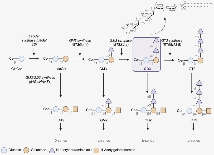
Fig.1 Ganglioside biosynthesis pathway highlighting the formation of GD3.1
The pathway explains why counter-screening cannot be treated as
an optional final check. GD3 is formed from GM3 and feeds structurally related downstream gangliosides. Testing
these neighbors early helps distinguish recognition of the intended disialylated arrangement from binding to a
shared precursor motif, lipid scaffold, or presentation artifact.
Project-Framing Questions
Project Question
Why It Matters
Which GD3 form should be recognized?
Defines whether the project focuses on canonical GD3, modified GD3 species, or a deliberately broader
recognition profile.
Where will the final reagent be used?
Determines whether purified lipid, liposome, cell, tissue, or more than one presentation format should
be included.
Which related gangliosides should be counter-screened?
Defines the scientific boundary of acceptable specificity before candidate selection becomes too
narrow.
What output is required?
Separates detection reagents, sequence-defined binders, and formats intended for downstream functional
research.
Comparator Panel and Interpretive Value
Comparator
Question Addressed
Interpretive Value
GD2
Does binding depend on the sialylation pattern that distinguishes GD3?
Tests discrimination from a close downstream ganglioside.
GM3
Is the antibody recognizing a shared precursor feature?
Reveals binding that may not require the full GD3 headgroup.
9-O-acetyl-GD3 or GD3 lactone
Do common GD3 modifications preserve or alter recognition?
Defines whether the reagent is canonical-GD3 selective or modification tolerant.
Lipid-, carrier-, or matrix-only controls
Is signal caused by the presentation system?
Identifies non-glycan and assay-format artifacts.
Our Anti-GD3 Antibody Development Services
Every anti-GD3 project starts from a different endpoint. Some
customers need a selective staining reagent for GD3-positive cells; others need a recombinant sequence for later
engineering or a panel that can compare canonical and modified GD3. Creative Biolabs brings antigen preparation,
antibody discovery, screening, and characterization into one coordinated program, while keeping the route
flexible enough to reflect the sample and assay that matter to the study.
GD3 Immunogen and Antibody Development
The antigen strategy may use glycan mimicry, GD3 conjugates, lipid-coated liposomes, or another reviewed
presentation. Discovery options described in our current service portfolio include hybridoma, phage display,
and single B-cell approaches.
Specificity and Cell-Surface Validation
Related gangliosides, lipid-only controls, carrier controls, and membrane-context assays can be combined
according to the antigen format. Candidates intended for cellular work can then be examined on relevant
GD3-positive and comparison models.
Membrane-Mimetic Design for GD3 Presentation
Conjugated or plate-bound GD3 offers controlled screening, while liposomal presentation restores a
hydrophobic environment and allows lipid composition and surface density to be considered deliberately.
Present the GD3 headgroup in a format suited to discovery and screening.
Antibody discovery
Hybridoma, phage display, or single B-cell routes
Match the discovery strategy to antibody format and downstream use.
Screening and counter-screening
GD3 with GD2, GM3, modified GD3, and format controls
Define the observed recognition profile and remove presentation-driven hits.
Characterization and validation
ELISA, flow cytometry, IHC, IF, TLC immunostaining, or other agreed assays
Confirm performance in the assay context relevant to the project.
Our Anti-GD3 Antibody Development Workflow
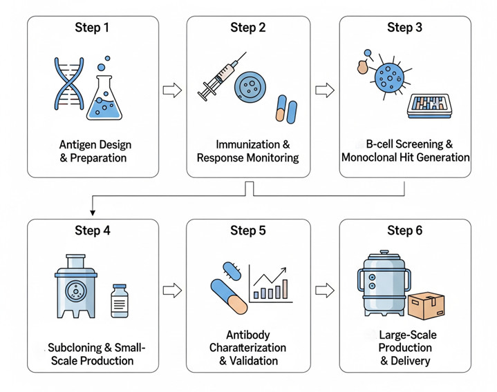
Our workflow moves from controlled antigen questions to the
more complex biological context. This staged approach preserves candidate diversity early, introduces
related-ganglioside selection before expensive downstream work, and gives the customer clear points at which to
review the data and refine the next step.
Fig.2 Custom anti-GD3 antibody development workflow.
Select a GD3 format that reflects the desired epitope and build in lipid, carrier, and
related-ganglioside controls.
Choose an antibody discovery route in view of GD3 immunogenicity, the desired format, and later
engineering needs.
Identify GD3-reactive candidates while removing lipid-only, carrier-only, and obvious cross-reactive
signals.
Confirm reproducible binding and retain candidates with distinct, interpretable recognition profiles.
Compare relevant gangliosides and test the shortlisted antibodies in the agreed membrane, cell, or
tissue context.
Prepare the selected research antibody format and associated project data according to the agreed
scope.
Project Requirements and Deliverables
A focused project discussion helps us distinguish essential
requirements from useful options. The deliverable package is then defined during scoping so conclusions remain
tied to the GD3 form, counter-target panel, antibody format, and assay conditions that were actually evaluated.
Project Input
Project Requirements
Share the intended GD3 form and the material available for antigen preparation or comparison.
Indicate whether the project should consider conjugates, liposomes, purified lipid, or another
format.
List GD2, GM3, modified GD3 species, or other scientifically relevant counter-targets.
Provide the planned cell, tissue, membrane, or assay context used for final evaluation.
Define whether the expected output is a detection reagent, sequence-defined binder, or recombinant
format.
Share existing antibodies, customer-supplied antigen, controls, or prior data when available.
Project Output
Deliverables and Validation Scope
Candidate antibodies selected within the agreed screening and specificity framework.
Sequence-defined or reformatted outputs when these are included in the agreed scope.
Data describing recognition across GD3, counter-targets, and relevant format controls.
Results from agreed cell, membrane, tissue, or assay-context confirmation.
Project data and conclusions tied to the GD3 form, comparator panel, format, and conditions that were
actually evaluated.
Discriminating GD3 from Related Gangliosides
Specificity is best understood as a measured recognition
profile, not as a label attached after one positive assay. A project may prioritize strict GD3 selectivity,
intentional recognition of a modified form, or a broader ganglioside pattern. The comparator panel should
reflect that goal and the biological system in which the antibody will be used.
Research Applications
Anti-GD3 antibodies can serve as research tools wherever GD3
abundance, accessibility, or molecular context needs to be measured. The required validation should follow the
application: an antibody selected for lipid profiling does not automatically qualify for intact-cell staining,
and a cell-binding reagent does not by itself demonstrate internalization or functional activity.
Cell Profiling
Profile GD3-positive populations by flow cytometry, immunofluorescence, or immunohistochemistry.
Comparative Ganglioside Studies
Compare canonical GD3 with GD2, GM3, or modified GD3 species in controlled models.
Mechanistic Research
Investigate GD3-associated adhesion, signaling, or immune-modulation hypotheses.
Sequence-Defined Formats
Develop Fab, scFv, or other research formats for downstream engineering studies.
All antibodies, services, and applications described here are
intended for research use only.
Start Your Anti-GD3 Antibody Development Project
Share the GD3 form, required counter-targets, intended assay, desired antibody format, and the validation
evidence needed. Creative Biolabs will use these parameters to discuss a project plan suited to your research
objective.
Discuss Your
Project
Published Data
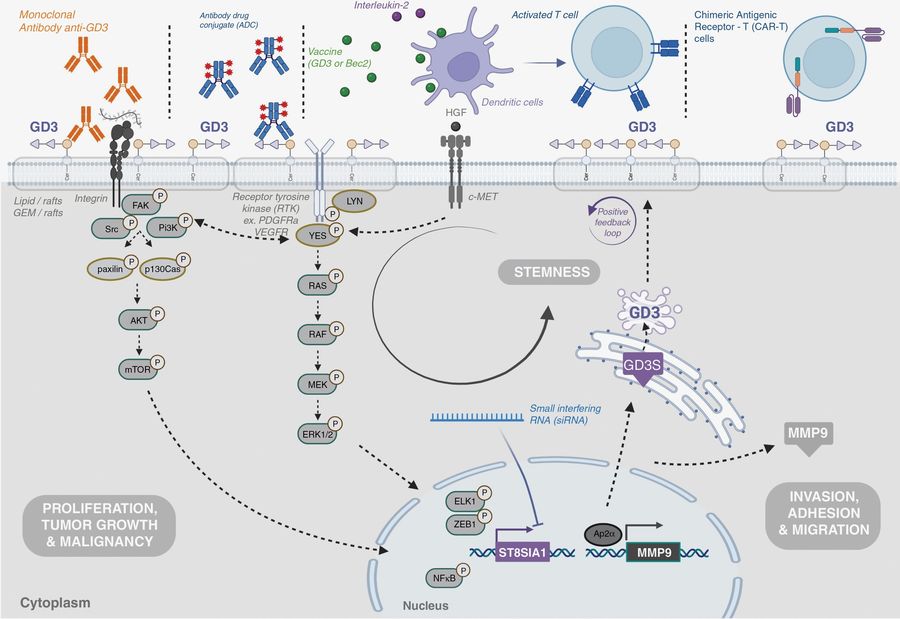
Fig.3 Overview of research strategies directed at GD3 and GD3 synthase.1
Cao and colleagues2 reviewed the biology of GD3, GD2, and GD3 synthase in cancer research, placing antibody-
and cell-based strategies within the wider landscape of GD3-directed investigation. The review highlights
both the biosynthetic relationship between gangliosides and the diversity of research formats used to study
them. For antibody development, this context reinforces the value of related-ganglioside counter-screening
and intact-cell confirmation.
Recommended Services or Products You May Need
Some projects need a custom discovery program; others are
better served by profiling an existing antibody or starting from a ready-to-use reagent. Use the tabs below to
move directly to the route that fits the next decision.
Product specifications and application listings should be
checked on the linked product pages when selecting a catalog reagent for a particular experiment.
Why should GD2 and GM3 be included in an anti-GD3 counter-screening panel?
GD2 and GM3 sit close to GD3 in the ganglioside biosynthetic
network and share structural features with it. Testing them helps determine whether a candidate recognizes the
complete GD3 headgroup or a common motif. Additional gangliosides can be added when the sample type or
research hypothesis makes them relevant.
Can the project distinguish canonical GD3 from 9-O-acetyl-GD3 or GD3 lactone?
Yes, when the required GD3 forms can be supplied or presented
in suitably matched formats. The comparison should control antigen density, lipid composition, and assay
geometry so that a signal difference can be attributed to the modification itself rather than to unequal
presentation or accessibility.
Is purified GD3 screening sufficient for an antibody intended for cell-surface studies?
Purified screening is useful for identifying and comparing
candidates, but it cannot reproduce every feature of a cell membrane. A cell-surface application should
include direct confirmation on an appropriate GD3-positive model together with a negative or low-expression
comparator and controls that help interpret target accessibility.
Which antibody discovery route is most appropriate for a GD3 project?
The choice depends on GD3 immunogenicity, the desired antibody
format, available antigen presentations, and whether sequence-defined outputs or later engineering are
important. Hybridoma, display-based, and single B-cell routes offer different advantages. Creative Biolabs
reviews these factors with the customer before recommending a project-specific strategy.
Can an existing anti-GD3 antibody be reformatted as a Fab, scFv, or another recombinant format?
A sequence-defined antibody can generally be evaluated for
recombinant reformatting. Because valency and architecture may change apparent avidity or membrane binding,
the reformatted molecule should be reassessed against GD3, relevant counter-targets, and the intended
biological presentation rather than assumed to retain the parent antibody's complete profile.
What information should we provide before requesting an anti-GD3 development proposal?
Please share the intended GD3 form, antigen source or
preferred presentation, required counter-targets, biological model, final assay, desired antibody format, and
any existing reagents or data. These details help define appropriate controls and avoid building the discovery
program around a presentation that does not match the downstream experiment.
References
Hein, Victoria, et al. "GD3 ganglioside is a promising therapeutic target for glioma patients." Neuro-Oncology Advances 6.1 (2024): vdae038. Distributed under Open Access license
CC BY 4.0, without modification. https://doi.org/10.1093/noajnl/vdae038
Cao, Shangqi, et al. “The Biological Role and Immunotherapy of Gangliosides and GD3 Synthase in Cancers.” Frontiers in Cell and Developmental Biology 11 (2023): 1076862. Distributed under Open Access license
CC BY 4.0, without modification. https://doi.org/10.3389/fcell.2023.1076862
Project Inquiry
!For Research Use
Only.Not for clinical or diagnostic use.