Creative Biolabs is offering the most comprehensive services for antibody development projects. With strict regulation and effective execution, we are dedicated to providing the most valuable solutions to complete your projects.
HighlightsWith over a decade of experience in phage display technology, Creative Biolabs can provide a series of antibody or peptide libraries that are available for licensing or direct screening. These ready-to-use libraries are invaluable resources for isolating target-specific binders for various research, diagnostic or therapeutic applications.
HighlightsCreative Biolabs has established a broad range of platforms for developing novel antibodies or equivalents. These cutting-edge technologies enable our scientists to meet your demands from different aspects and tailor the most appropriate solution that contributes to the success of your projects.
HighlightsWith deep understanding in antibody-related realms and extensive project experience, Creative Biolabs offers a variety of references to help you learn more about our capacities and achievements, including infographic, flyer, case study, peer-reviewed publications, and all kinds of knowledge that can assist your projects. You are also welcome to contact us directly for more specific solutions.
HighlightsGet a real taste of Creative Biolabs, one of the most professional custom service providers in the world. We are committed to providing highly customized comprehensive solutions with the best quality to advance your projects.
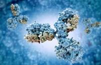
HighlightsSince the discovery of phage display technology, it has become an important research tool in biological research. Phage display technology fundamentally changes the process of traditional monoclonal antibody preparation, greatly reducing the time and complexity of the preparation. At present, this technology is widely used in the establishment of antigen and antibody libraries, drug design, vaccine research, pathogen detection, gene therapy, and many other fields. In addition, based on phage display technology, various methods, such as ribosome display and mRNA display, have been developed. Drawing upon our extensive practical experience and state-of-the-art technology platform, Creative Biolabs provides comprehensive development services for antibody phage display.
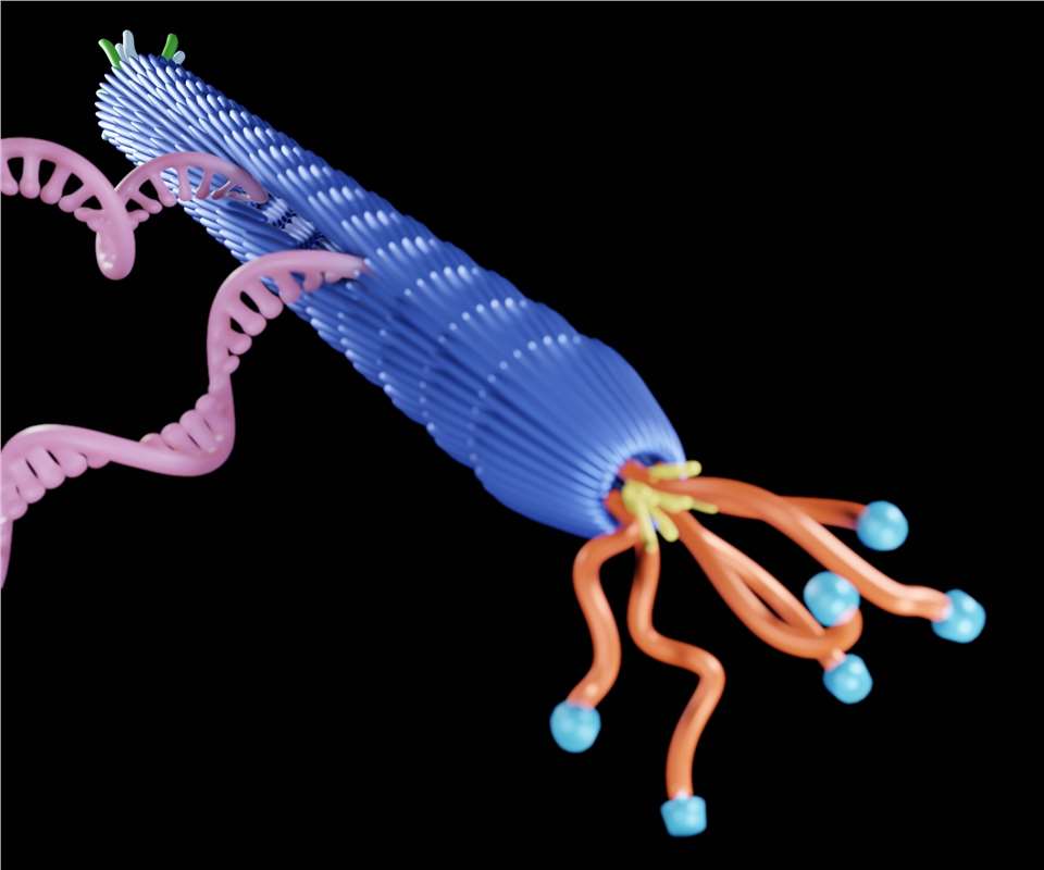
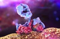
Phage display technology is to insert the gene encoding a foreign polypeptide or protein into the appropriate location with the phage coat protein gene using genetic engineering technology, and it can be correctly expressed in the reading frame so that the foreign polypeptide or protein forms a fusion protein on the capsid protein of the phage. These fusion proteins are presented on the phage surface with the reassembly of the phages and can maintain relative spatial structure and biological activity.
For high-affinity antibody screening using phage display technology, it is imperative to construct an antibody library first and then screen the phage library. The foreign gene is inserted into the phage gene, and as the phage passes through generations, the foreign protein is displayed on the surface of the daughter phage. This collection of bacteriophages displaying various foreign genes is known as the phage display library. Commonly used phage display library construction methods include the synthetic method, cDNA method, DNase method, etc.
Phage display library screening mainly involves two methods: in vivo screening and in vitro screening. In vivo screening refers to the intravenous injection of the phage display library into animals, leveraging the heterogeneity of the vascular molecular endothelium to selectively guide bacteriophages to different tissues. This leads to the acquisition of phage display antibodies specifically bound to different tissues. In vitro screening methods include biological elution, competition, reduction, selective infection screening, delayed infection screening, and other techniques, with biological elution being the most commonly used.
There are many classification criteria for phage display techniques. According to the different phages, they can be divided into M13, T7, T4, λ, and other phage systems. According to the different carriers, they can be divided into true phages and phagemids. According to the different libraries, it can be divided into random peptide libraries, cDNA libraries, antibody libraries, protein libraries, and other types. Each type of phage display technique has its own unique advantages and disadvantages. Therefore, when preparing premade antibody libraries, for example, selection and optimization should align with the specific experimental projects and research requirements.
Phage Display Systems Introduction
Phage Display Library Types Introduction
Phage Display Library Screening Strategy Introduction
Phage Display Applications Introduction
Creative Biolabs has a wealth of knowledge and experience in antibody phage display. We would be happy to discuss with you our knowledge and experience in phage display systems.
All listed services and products are For Research Use Only. Do Not use in any diagnostic or therapeutic applications.