3D Cell Culture
Creative Biolabs provides a full range of products and services based on our three-dimensional (3D) cell culture platform. With the well-established static and fluidic 3D cell culture systems, our services include, but are not limited to, establishment and optimization of experimental protocols, a series of in vitro analysis and screening (e.g., penetrability analysis, toxicity analysis and targeting analysis), as well as design custom in vitro 3D models (e.g., specific tumor tissue, in vitro organoid, and tissue-/organ-on-a-chip). Creative Biolabs is experienced and dedicated to helping customers in areas of 3D cell culture and analysis.
What is 3D cell culture?
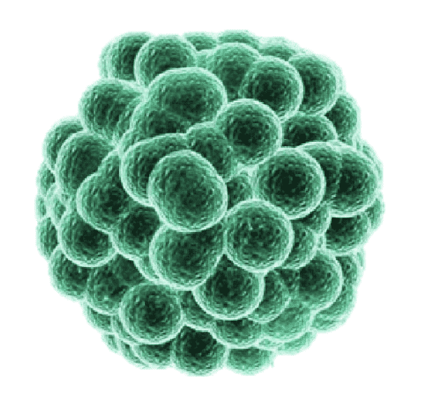
3D cell culture is a range of techniques that grow cells into 3D structures. Driven by the desire of biochemical and biomedical scientists for experimental systems that better represent the in vivo environments, new materials and methods for 3D cell culture has been developed. Application of 3D cell culture has resulted in better data and innovative knowledge of tissue and cancer behavior. Compared to two dimensional (2D) cell culture models, advantages of employing 3D cell culture models in the assessment of drug candidates can include: i) incorporated/increased cell-cell and cell-matrix interactions; ii) the impact of site specific stromal cells in tumor microenvironments can be evaluated; iii) varying cell proliferation zones; iv) oxygen, nutrient, metabolites, and catabolites gradients are established mimicking solid tumor environment; and v) the heterogeneous distribution of drug candidates that leads to the non-uniform exposure of cells to the tested molecules. Due to the variations of cell growth patterns, cell signaling pathways and target dependence differentiate between cells cultured in 2D and 3D structures. In vitro 3D cell culture models may provide more accurate insight into drug activity in vivo, which may not be thoroughly evaluated only based on 2D cell culture models. Currently existing 3D cell culture techniques can be grouped into two main categories, scaffold-based and scaffold-free 3D cell culture.
Technical basics
- Scaffold-based 3D cell culture
For the scaffold-based 3D cell culture, pre-fabricated scaffolds or matrices are designed to mimic the in vivo extracellular matrix (ECM). Upon introduction, cells attach, migrate, proliferate, and fill the interstices within the scaffold to form 3D structures. Scaffolds, also called 3D matrices, include a large variety of materials with diverse porosity, permeability, and mechanical characteristics. The scaffold and matrix can be derived from natural/biological origins or be synthesized via chemical reactions. Many common biomolecules are good candidates as 3D matrix/scaffold, including fibronectin, collagen, laminin, and gelatin…. Besides providing attachment sites and supports for cells, the natural/biological scaffold can also provide the correct microenvironment of growth factors, hormones, and other molecules. Those small molecules interact with cells and play crucial roles in gene expression and protein production. Scaffolds derived from natural resources can also promote appropriate cell behaviors and functions, resulting in increased cell viability, proliferation, and differentiation.
A wide range of synthetic materials can also serve as 3D matrix/scaffolds, including but not limited to polymers, ceramics, glass, carbon nanotubes, nanofibers, and various hybridizations. During the design and fabrication of scaffolds, a series of properties should be taken into consideration, including biocompatibility, wettability, mechanical property and structure, as well as surface chemistry. The high controllability and flexibility of synthetic scaffolds enable their wide applications. By providing a surface for cell growth and incorporating with essential nutrients and growth factors, they can easily impart and alter the 3D cell growth by minor alteration to the cell culture procedures.
- Scaffold-free 3D cell culture
For scaffold-free 3D cell culture, diverse technologies can be applied to achieve the cell culture in three dimensions. One of the most widely used strategies is the hanging drop plate (HDP). In the absence of a surface for attachment, cells will self assemble into 3D spheroid structures. HDP takes advantage of this fact by using well bottoms with an opening, which is different from traditional bottoms of culture plates. The aperture of the HDP bottom opening is carefully designed that is big enough to form a discrete media droplet sufficient for cellular aggregation, but also small enough to prevent the dislodgement of droplets during manipulation by taking advantage of surface tension. It takes from hours to days to form the final spheroid structure with size controlled by the initial number of cells seeded into the droplet. Co-cultured spheroid is feasible by using this HDP strategy.
Another type of plates suitable for the creation of tumor spheroid or multi-cell tissue models is microplates with a layer of ultra-low attachment (ULA) coating that minimizes cell adherence and allow spheroid formation. The round, tapered or v-shaped well bottoms of the ULA coated plates both ensure the generation of consistent, single spheroids, and facilitate in positioning spheroids in the middle of each well. Besides, 3D cell culture structures can be formed in microfluidic systems, where a perfused flow is introduced into the culture environment. The microfluidic system enables continuous introduction of nutrition and oxygen and in the meantime, ensures timely waste removal. With the introduction of a perfused flow as well as the incorporation of diverse physical and non-physical barriers, microfluidic 3D cell culture systems upgrade the complexity of culture models to a level much similar to that of the in vivo models.
Comprehensive 3D cell culture systems from Creative Biolabs
With years of research and development, Creative Biolabs is experienced in 3D cell culture techniques. By using either scaffold-based or scaffold-free 3D culture strategies, we can generate both mono-cell type and multi-cell type spheroid models for customers' specific demands. With well-established static and microfluidic 3D cell culture systems, our 3D cell culture platform fulfills requirements for generating custom in vitro 3D culture models, e.g., tumor tissues, micro-tissues, micro-organoids and tissue/organ-on-a-chip.
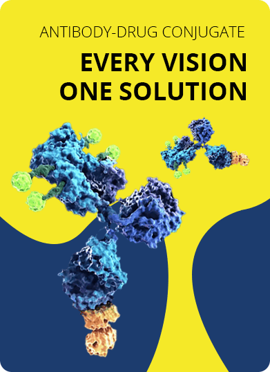
With expertise in biomaterial in vitro analysis, Creative Biolabs also provides a full range of in vitro analysis services to characterize the in vitro efficacy of antibody-drug conjugates (ADCs) based on our comprehensive 3D cell culture systems. Various parameters of ADCs can be evaluated using in vitro 3D culture models, including but not limited to distribution, tissue penetration, targeting efficacy and specificity, cytotoxicity, as well as bystander killing efficiency. Our scientific team provides customers with help from the experimental design to the establishment and optimization of protocols, collection and analysis of data, as well as suggestions on the improvement of the ADCs. Please contact us for more information and a discussion to see how we can be involved in your projects.
For Research Use Only. NOT FOR CLINICAL USE.
Online Inquiry
Welcome! For price inquiries, please feel free to contact us through the form on the left side. We will get back to you as soon as possible.
