
Choose the biological context
Define RPE source, differentiation stage, passage, plate or Transwell format, and baseline phenotype.
RPE secretome assay development
Measure how RPE models release pigment epithelium-derived factor in response to maturation, stress, and candidate compounds—with a study design built for meaningful conditioned-medium comparisons.
Start a PEDF Study Talk to a ScientistWhy measure secreted PEDF?
Pigment epithelium-derived factor is released by retinal pigment epithelium and is studied in retinal homeostasis, neuroprotection, and angiogenic balance. Quantifying PEDF in culture supernatant adds a functional layer to iPSC-facilitated AMD discovery that intracellular marker data alone cannot provide.
The assay is configured around a precise comparison rather than a generic protein measurement. Collection timing, medium volume, culture maturity, cell health, and normalization are aligned before execution.
A paired secretome perspective
The bars are conceptual, not performance data. A study can report PEDF alone or assess PEDF and VEGF as separate quantitative endpoints under matched sampling conditions.
Define RPE source, differentiation stage, passage, plate or Transwell format, and baseline phenotype.
Set test articles, vehicle controls, dose levels, exposure duration, and biological replication.
Fix the sampling window, volume, compartment, storage conditions, and normalization denominator.
Assay configuration
Creative Biolabs can integrate PEDF analysis into a customized stem cell-based assay development program. Each module is selected according to the model and the decision the data must support.
Measure PEDF in conditioned medium against a standard curve with study-specific sample dilution and technical replication.
Relate secretion to viable cells, total cellular protein, culture area, or collection duration to support between-group comparison.
Collect apical and basal compartments separately when a polarized Transwell RPE system is part of the agreed study.
Add VEGF, viability, morphology, or cell marker expression analysis where additional biological context is required.
Project workflow
Every stage has a defined checkpoint, so sample handling and normalization decisions are recorded before the final comparison is made.
Lock the model, groups, controls, replicates, collection interval, and reporting endpoint.
Checkpoint: approved study map
Prepare the RPE model, apply the agreed perturbation, and document culture condition and timing.
Checkpoint: treatment record
Harvest conditioned medium consistently and quantify PEDF with standards, controls, and replicates.
Checkpoint: accepted assay run
Apply the agreed denominator, compare groups, and integrate complementary endpoints where included.
Checkpoint: final data report
What you receive
The final package is configured during project scoping and can include raw, calculated, normalized, and visual outputs.
What we confirm
These inputs determine whether the resulting secretion values can be compared without avoidable ambiguity.
Research applications
The assay can be positioned as a compound-response endpoint, an RPE maturation measure, or one component of a broader secretome and phenotype strategy.
Compare PEDF release across disease-relevant RPE models, controls, and treatment conditions.
Evaluate dose- and time-dependent PEDF responses while controlling for cell viability.
Add a secretome endpoint to the characterization of RPE generated from selected iPSC lines.
Build inflammation, oxidative stress, polarity, or maturation studies around a defined PEDF endpoint.
Share the RPE model, treatment groups, collection window, and preferred normalization approach.
Open-access evidence
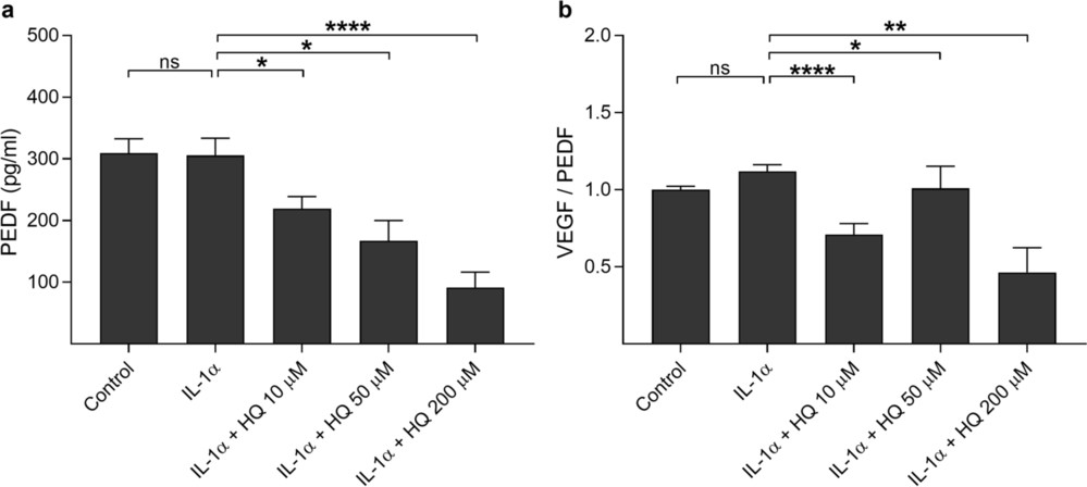
Bhattarai et al. primed ARPE-19 cells with IL-1α and then exposed them to 10, 50, or 200 μM hydroquinone for 18 hours. PEDF and VEGF were quantified in conditioned medium by ELISA. Hydroquinone significantly reduced extracellular PEDF at every tested concentration, with a concentration-dependent response; the lower concentrations affected secretion before overt loss of cell viability.
Assay relevance: The study demonstrates how PEDF secretion can be used as a treatment-sensitive endpoint in an RPE stress model and why paired viability and VEGF measurements strengthen interpretation.
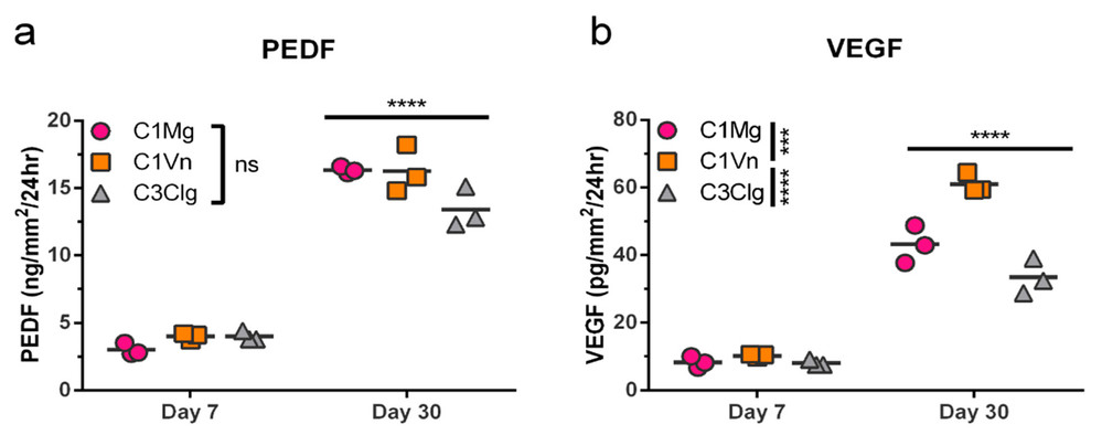
Faynus et al. cultured human pluripotent stem cell-derived RPE on three microcarrier systems and quantified PEDF in 24-hour conditioned-medium samples by sandwich ELISA. PEDF secretion increased significantly from day 7 to day 30 after seeding across all three microcarrier conditions, while no significant PEDF difference was detected between carrier types at the same time point.
Assay relevance: The results support longitudinal PEDF measurement as a functional maturation readout and illustrate the importance of matching collection duration and culture scale across groups.
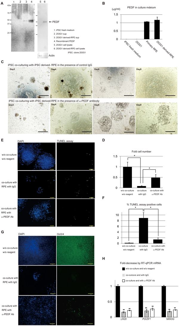
Kanemura et al. collected conditioned medium after 24 hours from primary RPE and 253G1 iPSC-derived RPE. ELISA detected more than 1 μg/mL PEDF in both RPE-conditioned media, and Western blotting confirmed the approximately 50 kDa PEDF protein. Neutralization experiments further linked secreted PEDF to the observed effect on residual iPSC growth.
Assay relevance: This work illustrates quantitative PEDF measurement in iPSC-RPE supernatant, orthogonal protein confirmation, and the use of a functional experiment to investigate the biological meaning of the secreted factor.
Frequently asked questions
The assay quantifies pigment epithelium-derived factor released into conditioned culture medium. Results can be reported as concentration, normalized secretion, or fold change relative to a control condition.
Projects may use conditioned medium collected from RPE cells, iPSC-derived RPE cultures, or another agreed research cell model. Medium composition, collection timing, storage, expected range, and sample volume are reviewed before testing.
Yes. A paired design can quantify both factors when the study requires evaluation of anti-angiogenic and pro-angiogenic balance. The appropriate interpretation depends on the model, treatment, viability, and normalization strategy.
Depending on the culture system, PEDF may be normalized to viable cell number, total cellular protein, culture area, collection duration, or another agreed denominator. The same collection and normalization method should be applied across comparison groups.
No. This service is for research use only and is not intended for diagnostic or clinical use.