
Flow Cytometry
Quantify the proportion of PMEL-positive cells and compare fluorescence distributions across experimental groups using a predefined gating and control strategy.
Marker-focused assay development
A customizable research workflow for detecting and quantifying PMEL/PMEL17 expression in stem-cell-derived pigment cell models, with readouts selected around your sample type and scientific question.
Discuss Your Assay Request Assay ConsultationBiological context
PMEL, historically referred to as PMEL17 or gp100, is a melanosome-enriched protein involved in the structural organization of premelanosomes. Its expression is commonly examined in melanocytic and retinal pigment epithelium (RPE) research, including iPSC-facilitated discovery for age-related macular degeneration.
Because a single marker does not define cell identity or functional maturity by itself, Creative Biolabs structures PMEL17 analysis around the experimental context. PMEL results can be interpreted alongside morphology, complementary lineage markers, pluripotency marker expression, and other project-specific endpoints.
Modular assay options
A project may use one method or an orthogonal combination within a customized assay development plan. Final platform selection depends on sample format, available material, desired level of quantitation, and the biological endpoint.
Quantify the proportion of PMEL-positive cells and compare fluorescence distributions across experimental groups using a predefined gating and control strategy.
Visualize PMEL expression and intracellular localization while preserving cellular morphology and enabling co-staining with selected companion markers.
Measure relative PMEL transcript abundance with project-defined normalization genes, controls, and comparison groups.
Assess PMEL protein signal in cell lysates and compare relative abundance across conditions with an appropriate loading-control strategy.
Project workflow
The workflow is scoped around explicit sample, control, and reporting requirements.
Define the model, comparison, endpoint, and success criteria.
Select the platform, controls, replicates, markers, and sampling points.
Confirm sample compatibility and optimize critical detection conditions as needed.
Run the agreed assay with documented sample and control handling.
Deliver analyzed results, method details, and project-specific interpretation.
Outputs and planning
Available outputs and required inputs are confirmed in the study plan before execution.
| Project Input | Details to Confirm |
|---|---|
| Biological model | Cell type, species, differentiation stage, culture format, and experimental treatment. |
| Sample format | Live or fixed cells, slides, lysate, RNA, or another agreed research sample. |
| Comparison design | Reference condition, controls, sample groups, replicates, and collection time points. |
| Readout | Population percentage, fluorescence intensity, localization, relative RNA expression, or protein abundance. |
| Reporting needs | Required figures, data format, statistical summary, and documentation expectations. |
Research applications
PMEL analysis is most informative when it is integrated into a defined experimental framework and interpreted with complementary evidence.
Track PMEL expression as one component of a broader marker strategy for stem-cell-derived RPE models in AMD research.
Compare PMEL expression across melanocytic differentiation conditions or experimental groups generated from iPSCs.
Evaluate how changes in culture conditions, timing, or candidate factors affect a selected PMEL endpoint in a drug discovery workflow.
Add an RNA- or protein-level PMEL readout to a multiparameter cell identity and phenotype assessment.
Open-access evidence
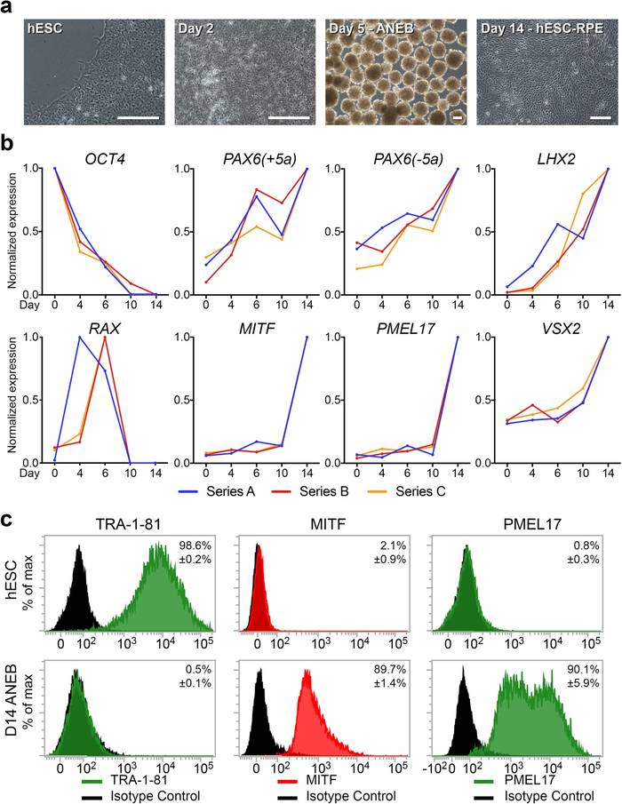
Limnios et al. evaluated human embryonic stem cell-derived RPE generated under defined conditions using a panel that included PMEL17, MITF, and the pluripotency marker TRA-1-81. Flow cytometry detected PMEL17 in approximately 85% of cells at day 14. Following extended expansion, passage 8 cultures retained PMEL17 with other RPE-associated markers, while TRA-1-81 remained undetectable.
Assay relevance: The study supports using PMEL17 within a multiparameter panel to assess RPE differentiation and phenotype maintenance across culture stages.
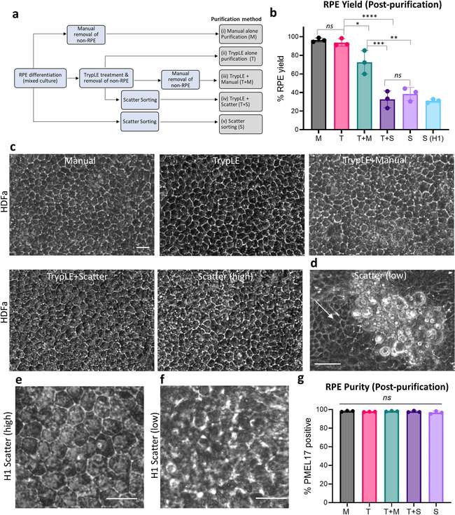
Regha et al. compared five purification approaches for RPE cultures derived from different stem cell sources. The investigators used PMEL17 flow cytometry to estimate post-purification purity, RT-qPCR for PMEL17 and RPE65 to distinguish cell fractions, and PMEL17/ZO-1 immunostaining to assess phenotype. All five strategies produced populations with more than 96.8% PMEL17-positive cells, although cell yield and processing time varied between methods.
Assay relevance: These results demonstrate how orthogonal PMEL17 readouts can support enrichment assessment, method comparison, and RPE process optimization.
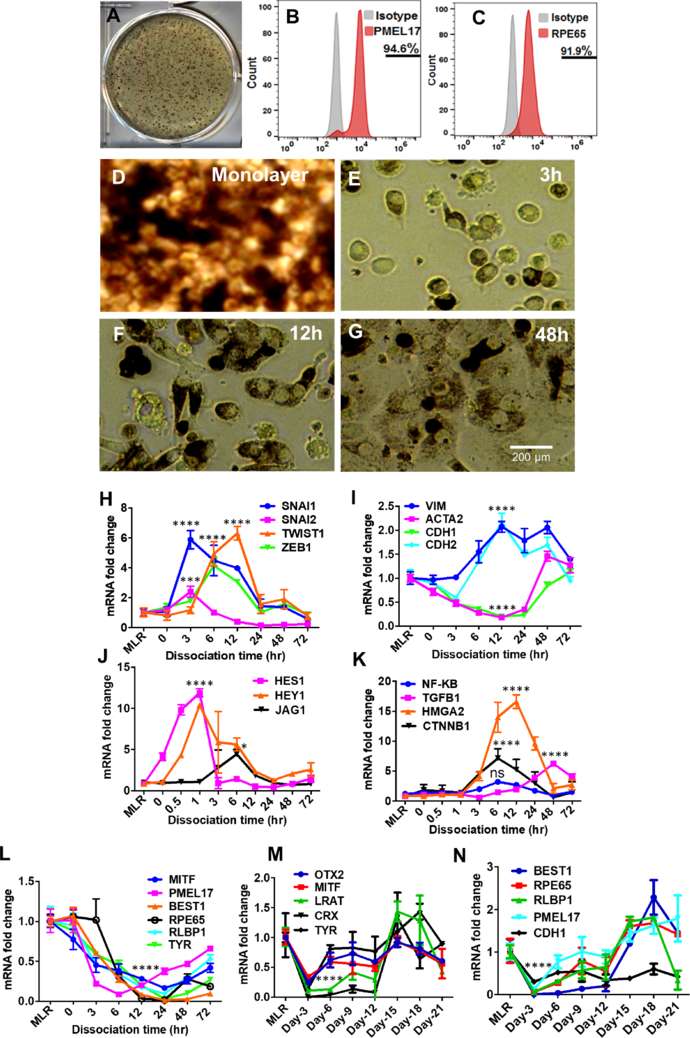
Sripathi et al. induced epithelial-to-mesenchymal transition in human stem cell-derived RPE using two complementary systems and followed the response by time-course RNA sequencing, RT-qPCR, and immunostaining. PMEL17 expression decreased approximately 12-fold as the RPE cultures transitioned toward a mesenchymal state.
Assay relevance: The work shows how PMEL17 can contribute to monitoring phenotype stability or loss when interpreted alongside epithelial, mesenchymal, and other RPE differentiation markers.
Frequently asked questions
A PMEL17 expression assay measures PMEL at the RNA or protein level. Depending on the project, it can report population-level expression, cellular localization, relative transcript abundance, or protein abundance.
Projects may begin with client-provided cells, lysates, RNA, fixed samples, or a stem-cell-derived pigment cell model. Sample compatibility, quantity, shipping, and handling requirements are confirmed during project design.
Yes. A project-specific panel can pair PMEL with markers selected for the intended lineage, maturation stage, pluripotency status, or experimental control strategy. The appropriate panel depends on the model and the claim the data must support.
PMEL expression should be interpreted in the context of the model, control conditions, assay platform, and companion evidence. PMEL positivity alone does not establish complete cell identity or functional maturity.
No. This service is for research use only and is not intended for diagnostic or clinical use.