

Introduction to iPSC-Derived Ocular Cell Models
Induced pluripotent stem cells provide a powerful starting material for generating human ocular cell types that are otherwise difficult to obtain in sufficient quantity, genetic diversity, or disease-relevant condition. Primary human retinal, optic nerve, and ocular surface cells are limited in availability and often unsuitable for longitudinal studies or high-throughput assays. In contrast, iPSCs can be expanded, genetically characterized, edited, banked, and differentiated into multiple ocular lineages under controlled in vitro conditions.
Ocular differentiation from iPSCs typically follows developmental logic. Pluripotent cells are guided toward neuroectodermal, surface ectodermal, optic vesicle-like, retinal progenitor, or ocular surface epithelial states through carefully timed modulation of signaling pathways such as BMP, WNT, TGF-β, IGF, FGF, Notch, and retinoic acid pathways. Depending on the project goal, cells may be differentiated in two-dimensional monolayer formats, three-dimensional organoid systems, co-culture platforms, maturation-enhanced formats, or assay-ready multiwell configurations.
The diversity of iPSC-derived ocular models enables researchers to select the most appropriate biological system for their question. Creative Biolabs develops ocular cell differentiation workflows that balance biological relevance, reproducibility, scalability, and practical assay compatibility. Our services can be integrated with upstream iPSC generation, genome editing, disease-specific iPSC banking, clone selection, and downstream phenotypic assay development.
Our Ocular Cell Differentiation Service Portfolio
Creative Biolabs provides a broad portfolio of iPSC-derived ocular cell differentiation services. Each service can be delivered as a stand-alone project or incorporated into a larger ophthalmology research program.
| Service Module | Main Deliverables | Typical Research Use |
|---|---|---|
| iPSC-RPE Differentiation |
|
AMD modeling, phagocytosis assays, barrier function, toxicity testing |
| Retinal Organoid Generation |
|
Retinal development, inherited retinal disease modeling, gene therapy evaluation |
| Photoreceptor-Like Cell Differentiation |
|
Retinal dystrophy studies, photoreceptor rescue assays, gene correction evaluation |
| Retinal Ganglion Cell Differentiation |
|
Glaucoma models, optic neuropathy, neuroprotection assays |
| Corneal Epithelial-Like Cell Differentiation |
|
Corneal injury models, ocular irritation, epithelial repair studies |
| Ocular Surface Lineage Development |
|
Dry eye, inflammation, mucosal barrier studies |
| Custom Ocular Differentiation |
|
Advanced therapeutic development, assay customization, translational studies |
Our team can start from client-provided iPSC lines, disease-specific iPSC clones, gene-edited iPSCs, or iPSC lines generated by Creative Biolabs. For projects involving rare disease samples, complex genotypes, or demanding downstream assays, we can perform feasibility assessment before launching full-scale differentiation.
Technical Workflow for iPSC-Derived Ocular Cell Differentiation
Ocular differentiation projects require careful control at every stage. Small differences in iPSC quality, passage number, colony morphology, seeding density, extracellular matrix, medium composition, timing of pathway modulation, and maturation strategy may influence final cell identity and assay performance. Creative Biolabs follows a structured workflow designed to reduce variability while preserving flexibility for custom development.
-
Project Design and Feasibility Assessment
Every project begins with a detailed discussion of scientific goals, disease context, required cell type, target markers, desired maturity level, assay format, scale, and data package. Our scientists evaluate whether the requested ocular lineage is best generated as a monolayer, organoid, enriched progenitor population, mature-like culture, co-culture, or cryopreserved intermediate. -
iPSC Line Evaluation and Pre-Differentiation QC
Before differentiation, iPSC lines are assessed for suitability. A high-quality starting population is essential for ocular lineage commitment. Creative Biolabs can evaluate morphology, viability, growth characteristics, mycoplasma status, pluripotency marker expression, karyotype stability, and differentiation readiness. -
Lineage Induction
The induction phase guides iPSCs away from pluripotency and toward ocular-relevant developmental programs. Depending on the target cell type, cells may be directed toward anterior neuroectoderm, eye field progenitors, optic vesicle-like structures, retinal progenitors, surface ectoderm, or epithelial progenitor states. -
Expansion, Enrichment, and Maturation
After initial specification, target ocular cells are expanded or matured according to project needs. For iPSC-RPE, maturation may include development of polygonal morphology, pigmentation, tight junction formation, and epithelial barrier-associated features. For retinal organoids, maturation may involve maintenance of laminated structures and generation of retinal cell populations over extended timelines. For RGC cultures, maturation may be evaluated through neuronal morphology, RGC marker expression, neurite extension, and functional assay compatibility. -
Characterization and Release Testing
Creative Biolabs provides tiered QC packages ranging from essential marker confirmation to in-depth molecular and functional characterization. Each project can be released with a documentation package aligned with client requirements. -
Delivery and Downstream Support
Final deliverables may include live cultures, cryopreserved cells, fixed plates, organoids at specified stages, lysates, RNA, images, reports, protocols, and assay recommendations. Our scientists can also assist with downstream assay design, troubleshooting, and scale-up planning.
Supported Ocular Cell Types
| Ocular Cell Types | Descriptions |
|---|---|
| iPSC-Derived Retinal Pigment Epithelium Cells |
Creative Biolabs can generate iPSC-RPE cultures with characteristic pigmentation, epithelial morphology, and RPE marker expression. Depending on project needs, we can provide early-stage RPE progenitors, mature RPE monolayers, cryopreserved RPE cells, or assay-ready RPE plates. Typical markers and readouts may include:
|
| iPSC-Derived Retinal Organoids | They may contain retinal progenitors and differentiated retinal cell types, including photoreceptor-like cells, ganglion cell-like populations, bipolar-like cells, amacrine-like cells, horizontal-like cells, and Müller glia-like cells depending on maturation stage and protocol. |
| iPSC-Derived Photoreceptor-Like Cells | Creative Biolabs supports photoreceptor-directed differentiation. Depending on the experimental design, photoreceptor-like cells may be generated within retinal organoids or as enriched populations. Characterization can include expression of photoreceptor-associated markers such as CRX, NRL, RCVRN, OTX2, ARR3, RHO, OPN1SW, OPN1MW/LW, and other project-specific targets. |
| iPSC-Derived Retinal Ganglion Cells | Creative Biolabs can develop RGC-enriched differentiation workflows using stepwise neural induction and retinal ganglion cell specification. RGC models may be delivered as neuronal cultures, fixed assay plates, or live cells for downstream functional testing. |
| iPSC-Derived Corneal Epithelial-Like Cells |
Creative Biolabs can provide differentiation strategies that direct iPSCs toward surface ectodermal and corneal epithelial-like identities. Depending on project requirements, cultures may be optimized for monolayer assays, stratified epithelial-like morphology, barrier function, or inflammatory response studies. Typical markers may include:
|
| Customized Ocular Lineage Systems |
Some projects require more specialized ocular models than standard RPE, retinal organoid, or RGC workflows. Creative Biolabs can design custom differentiation systems for:
|
Customization Options
Creative Biolabs understands that ocular research projects vary widely in biological complexity and practical requirements. Our iPSC-derived ocular differentiation services can be customized at multiple levels.
- Starting Material Options - We can work with client-provided iPSC lines, healthy donor iPSC lines, patient-derived iPSC lines, disease-specific iPSC panels, gene-edited iPSC clones, isogenic control pairs, reporter iPSC lines, and iPSC lines generated by Creative Biolabs.
- Differentiation Format Options - Available formats may include 2D monolayer cultures, 3D retinal organoids, ocular epithelial monolayers, assay-ready plates, long-term maturation cultures, cryopreserved intermediate cells, co-culture-ready populations, fixed imaging plates, and RNA or protein endpoint samples.
- Marker Panel Customization - Clients can request standard or customized marker panels.
- Functional Assay Integration - Creative Biolabs can help integrate differentiation with downstream assays.
- Timeline and Maturation Customization - Ocular cell differentiation timelines can vary considerably. RPE differentiation may be shorter than long-term retinal organoid maturation. Photoreceptor maturation and retinal organoid development may require extended culture. Creative Biolabs can align project timelines with the desired endpoint and provide staged deliverables when needed.
Published Data
iPSCs are emerging as a valuable system for modeling tissues and organs. The researchers described a highly scalable protocol for the differentiation of iPSCs into retinal pigment epithelium (RPE), including a new step that makes it easier for researchers to obtain a high-purity culture. They also describe a cryopreservation technique for RPE progenitor cells to enable their storage. The use of cryopreserved cells allows a subsequent reduction in differentiation time compared to the full protocol.
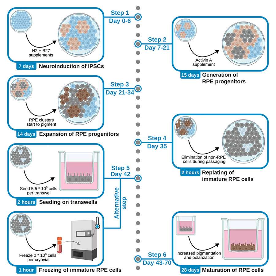 Fig. 1 A highly scalable protocol for the differentiation of iPSCs into retinal pigment epithelium (RPE).1,3
Three-dimensional retinal organoids (3D-retinas) are a promising graft source for transplantation therapy. The researchers previously developed self-organizing culture for 3D-retina generation from human pluripotent stem cells (hPSCs). Here they presented a quality control method and preclinical studies for tissue-sheet transplantation. Self-organizing hPSCs differentiated into both retinal and off-target tissues. Gene expression analyses identified the major off-target tissues as eye-related, cortex-like, and spinal cord-like tissues.
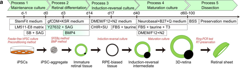 Fig. 2 Self-organizing culture of human iPSCs to generate the 3D-retina and dissected retinal sheet.2,3
What Our Clients Say
"Creative Biolabs helped us establish a patient-specific iPSC-RPE model for a macular degeneration program. The differentiated cells showed stable pigmentation, epithelial-like morphology, and strong expression of RPE-associated markers. The project team also provided clear guidance on culture handling and downstream assay planning, which allowed us to move smoothly into functional testing."
— Senior Scientist, Ophthalmology Discovery Group
"Our internal retinal organoid workflow was difficult to reproduce across multiple iPSC lines. Creative Biolabs helped us optimize the differentiation process, define realistic time points, and generate organoids with the developmental stage we needed for our inherited retinal disease study. The regular milestone updates were especially valuable."
— Principal Investigator, Academic Vision Research Center
"We approached Creative Biolabs for retinal ganglion cell differentiation to support a glaucoma-related neuroprotection project. The team understood that marker expression alone would not be enough for our goals. They helped us design a workflow that included neuronal morphology evaluation, neurite outgrowth analysis, and assay-compatible delivery."
— Translational Research Lead, Biotech Company
"The flexibility of the service was a major advantage. Our project involved gene-edited iPSC clones, isogenic control lines, ocular differentiation, imaging endpoints, and RNA sample collection. Creative Biolabs coordinated these steps as a single integrated workflow, which saved our team significant time."
— Director of Preclinical Research, Gene Therapy Company
FAQs
Q: What types of iPSC-derived ocular cells can Creative Biolabs generate?
A: Creative Biolabs can support the generation of multiple ocular cell types and ocular lineage models, including iPSC-derived retinal pigment epithelium cells, retinal organoids, photoreceptor-like cells, retinal ganglion cells, retinal progenitor-like cells, corneal epithelial-like cells, conjunctival-like cells, limbal epithelial-like cells, and customized ocular differentiation systems. The final cell type selection depends on your disease area, experimental objective, assay format, and required maturity level.
Q: Can you provide iPSC generation before ocular differentiation?
A: Yes. If you have donor somatic cells but not established iPSC lines, Creative Biolabs can support upstream iPSC generation, clone expansion, characterization, banking, and subsequent ocular differentiation. This integrated workflow is useful for projects that require patient-specific disease models or custom iPSC-derived ocular cell systems from the beginning.
Q: What starting materials can be used for this service?
A: Depending on the project, starting materials may include established iPSC lines, patient-derived iPSC lines, healthy donor iPSC lines, gene-edited iPSC clones, reporter iPSC lines, isogenic control pairs, or somatic cells for upstream reprogramming. Before project initiation, Creative Biolabs will review the available material and recommend the most suitable workflow.
Q: How long does iPSC-derived ocular cell differentiation take?
A: The timeline varies by target cell type and endpoint. iPSC-RPE differentiation is generally different from long-term retinal organoid culture, photoreceptor maturation, or retinal ganglion cell differentiation. Some applications require early progenitor-like cells, while others require more mature cultures or extended organoid development.
Q: What markers are commonly used for photoreceptor-like cells?
A: Photoreceptor-like cells may be characterized using markers such as CRX, OTX2, NRL, RCVRN, RHO, ARR3, OPN1SW, OPN1MW/LW, and other rod- or cone-associated markers. The selected marker panel depends on whether the project is focused on general photoreceptor development, rod-like populations, cone-like populations, or disease-specific phenotypes.
Q: Can Creative Biolabs provide assay-ready ocular cells?
A: Yes. For selected ocular cell types and applications, Creative Biolabs can provide assay-ready cells in multiwell plates, fixed imaging plates, live cultures, cryopreserved cells, organoids, RNA samples, protein lysates, or other customized delivery formats.
Q: Can you integrate genome editing with ocular cell differentiation?
A: Yes. Genome editing can be integrated upstream of ocular differentiation. Creative Biolabs can support mutant iPSC generation, gene correction, reporter line development, clone screening, genotype confirmation, and subsequent differentiation into relevant ocular cell types.
Take the Next Step with Creative Biolabs







1. Contact Us
via the Inquiry Form or Email
2. Define Your Needs
Cell Type, Function, Quantity, Modifications
3. Kickstart the Project
Our Expert Team Guiding Every Step
Ocular disease research requires models that reflect human biology, disease background, and experimentally meaningful cellular function. Creative Biolabs helps researchers access customized iPSC-derived ocular cell systems without the burden of building every differentiation workflow internally.
Contact Creative Biolabs today to discuss your iPSC-derived ocular cell differentiation project.
References
- Zorin, Mark, Rike Ewerling-Hähnel, and Kerstin Nagel-Wolfrum. "Protocol for the differentiation of induced pluripotent stem cells into retinal pigment epithelial cells." STAR protocols 6.4 (2025). https://doi.org/10.1016/j.xpro.2025.104263
- Watari, Kenji, et al. "Self-organization, quality control, and preclinical studies of human iPSC-derived retinal sheets for tissue-transplantation therapy." Communications Biology 6.1 (2023): 164. https://doi.org/10.1038/s42003-023-04543-5
- Distributed under Open Access license CC BY 4.0, without modification.