Pluripotency marker assays are critical for regenerative medicine, ensuring iPSC trilineage differentiation capacity and mitigating teratoma risks. Creative Biolabs' service uses multi-omic validation, flow cytometry, and immunocytochemistry to deliver quantified marker data. It meets gold standards, streamlines IND paths, and provides definitive project guidance for reliable, compliant iPSC characterization.
Discover How We Can Help - Request a Consultation
Pluripotency marker assay of induced pluripotent stem cells (iPSCs) is a core quality control method for verifying the pluripotent state of iPSCs. Through a multi-dimensional experimental strategy, it accurately evaluates the undifferentiated characteristics, self-renewal capacity, and differentiation potential of cells, providing critical data support for iPSC line establishment, subculture, and application.
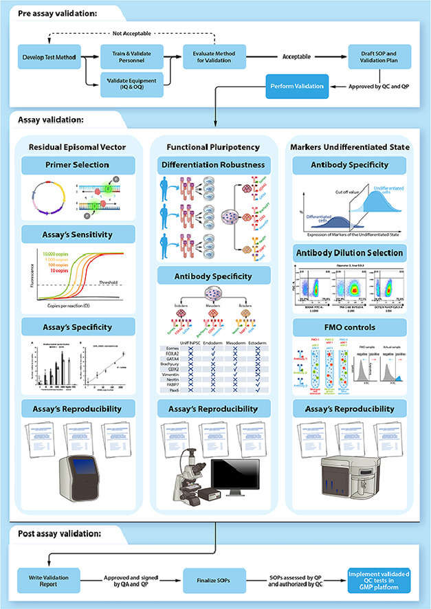 Fig.1 Utilize multiple methods to identify the pluripotency of iPSCs.1,3
Focusing on core transcription factors and specific genes that regulate stem cell pluripotency, common detection targets include:
| Detection Method | Core Advantages | Application Scenarios |
|---|---|---|
| qPCR/ddPCR | High quantitative accuracy and sensitivity, capable of detecting 0.001% of residual pluripotent cells | Quantification of gene expression levels, quality control screening |
| Immunofluorescence Staining | Direct visualization of protein expression and spatial distribution of cellular pluripotency | Single clone selection, morphological validation |
| Flow Cytometry | High-throughput detection, enabling simultaneous analysis of multiple surface markers | Phenotypic identification of large-scale cell populations |
| Western Blot | Verification of protein molecular weight and specificity | Qualitative/semi-quantitative analysis at the protein level |
Our standardized workflow is designed for maximum transparency and scientific rigor, transitioning seamlessly from sample receipt to a detailed analytical dossier.
As a global leader in stem cell solutions, Creative Biolabs provides highly customized Pluripotency Marker Analysis Service via Immunofluorescence (IF) Assay tailored to the specific genetic background and therapeutic goals of your cell lines. We don't just provide data; we provide a biological roadmap for your project. Our offerings include:
from initial reprogramming validation to pilot and large-scale bank certification.
tailored to specific cell types, allowing for the addition of niche markers or tissue-specific lineage indicators.
principles to ensure your pluripotency data meets stringent regulatory expectations for IND filings.
including simultaneous assessment of surface markers, intracellular proteins, and mRNA expression profiles.
are maintained throughout the cell expansion and analytical phases to prevent cross-contamination.
for specific clones to maximize the expression of key pluripotency factors before differentiation.
of cell origin and passage history, approved by our internal qualified quality assurance service.
Experience the Creative Biolabs Advantage - Get a Quote Today
Induced pluripotent stem cell lines (iPSCs) are generated from human peripheral blood mononuclear cells (PBMCs) via adenovirus-mediated reprogramming. On the 9th day post-reprogramming, the cells form tightly packed, dome-shaped colonies with distinct boundaries, and their morphology remains stable following 5 passages. With GAPDH as the internal reference gene, quantitative PCR analysis of the pluripotency markers OCT4, SOX2, NANOG, and KLF4 revealed that their expression levels in the generated iPSCs were comparable to those of embryonic stem cells. Furthermore, multiplex immunofluorescence staining and flow cytometry assays were performed to confirm the co-expression of pluripotency and proliferation markers in these cells.
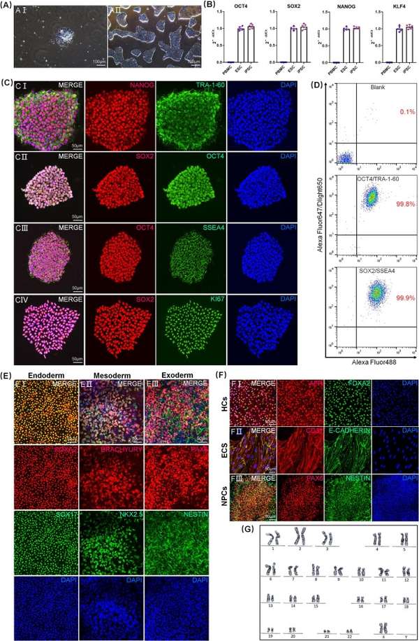 Fig.2 The pluripotency of iPSCs derived from peripheral blood mononuclear cells (PBMCs) was verified using techniques such as flow cytometry, immunofluorescence, and qPCR.2,3
A: We utilize a combination of surface markers (TRA-1-60) and core transcription factors (NANOG). Partial clones often express early markers like SSEA-4 but fail to activate the endogenous NANOG circuitry required for stable pluripotency.
A: While this specific panel is optimized for iPSCs/ESCs, we offer specialized mesenchymal stem cell (MSC) characterization panels following ISCT guidelines, including CD73, CD90, and CD105 markers.
A: Our high-sensitivity Flow Cytometry protocols can detect undifferentiated cells at a level of 0.01% or lower, which is critical for safety assessments in cell therapy products.
A: Yes, we strongly recommend pairing marker assays with karyotyping or G-banding to ensure that the reprogramming process has not introduced chromosomal abnormalities.
A: All results are compiled into a formal technical report that includes methodology, raw data, representative images, and a summary of findings aligned with GLP-like standards.
Creative Biolabs offers an end-to-end solution for Pluripotency Marker Analysis Service via Immunofluorescence (IF) Assay, providing the precision, scalability, and documentation necessary for modern regenerative medicine. From early-stage discovery to large-scale allogeneic manufacturing, our expertise ensures your cell lines are validated, stable, and ready for clinical application.
Contact Our Team for More Information and to Discuss Your Project
References
For Research Use Only. Not For Clinical Use.