
Why Use iPSC-Derived Osteogenic Cells?
Bone biology is shaped by complex interactions among osteoprogenitors, osteoblasts, osteocytes, osteoclasts, immune cells, vascular cells, extracellular matrix, and mechanical signals. Although no in vitro system can fully reproduce the native bone microenvironment, iPSC-derived osteogenic cells provide an adaptable and human-relevant model for studying many aspects of osteogenesis.
Primary human osteoblasts remain useful but are constrained by tissue availability, donor variability, limited expansion, and inconsistent maturation. Immortalized cell lines offer convenience but may not reflect normal differentiation behavior. Animal-derived systems can provide mechanistic insights, but species-specific differences may limit translation. iPSC-based osteogenic differentiation helps overcome some of these limitations by enabling researchers to work with renewable, genetically defined, and potentially patient-specific human cell sources.
iPSC-derived osteogenic cells are particularly valuable when researchers need:
1. Genetically consistent cell material
A single iPSC clone can be expanded and differentiated across multiple batches, supporting repeatable experimental designs and longitudinal studies.
2. Patient-specific or disease-specific bone models
iPSCs derived from patients with skeletal disorders can be differentiated into osteogenic cells to study disease-associated defects in commitment, matrix formation, mineralization, or response to treatment.
3. Isogenic comparison systems
Gene editing can be used to introduce or correct mutations in iPSCs, creating matched control and disease lines that differ only at defined genetic loci.
4. Scalable cell supply
iPSC expansion provides a renewable upstream source, making it possible to support screening-scale or multi-condition experiments.
5. Compatibility with advanced platforms
Osteogenic cells derived from iPSCs can be incorporated into 2D assays, 3D scaffolds, organoid-like systems, microfluidic devices, and co-culture models.
6. Controlled differentiation windows
Cells can be collected at defined stages, including mesenchymal-like progenitor, osteoprogenitor, pre-osteoblast, and mature osteoblast-like states.
Service Overview
Our iPSC-derived osteogenic cell differentiation platform is built to bridge the gap between pluripotent stem cell technology and practical bone research models. We support projects starting from client-supplied iPSC lines, internally generated iPSCs, disease-specific iPSCs, genome-edited iPSC clones, or validated control lines. Based on project needs, we can develop osteogenic differentiation workflows that emphasize reproducibility, lineage purity, maturation level, phenotypic stability, or compatibility with functional assays.
Our service may include:
- iPSC line recovery, expansion, and pre-differentiation quality review
- Directed differentiation toward mesenchymal and osteogenic lineages
- Generation of osteogenic progenitors, pre-osteoblasts, or mature osteoblast-like cells
- Osteogenic induction and matrix mineralization culture
- Morphological monitoring and differentiation-stage documentation
- Marker analysis by qPCR, immunostaining, flow cytometry, or Western blot
- Functional assays such as alkaline phosphatase activity, calcium deposition, and mineralized nodule formation
- Custom model development for skeletal disorders, bone toxicity testing, and regenerative medicine research
- Cryopreserved or live-cell delivery depending on project design
- Final technical report with methods, images, QC data, and recommended handling notes
Rather than offering a rigid one-size-fits-all protocol, Creative Biolabs develops practical osteogenic differentiation plans that match each client's biological question. For example, a project focused on early osteoblast commitment may require strong expression of RUNX2 and SP7, while a mineralization study may prioritize ALP activity, collagen matrix deposition, osteocalcin expression, and Alizarin Red-positive calcium accumulation. A biomaterial project may require cells adapted for 3D scaffolds, hydrogels, or coated surfaces. A disease modeling project may require matched control and mutant iPSC lines differentiated in parallel under tightly controlled conditions.
Our iPSC-Derived Osteogenic Differentiation Strategy
Creative Biolabs applies a staged differentiation strategy that guides iPSCs from pluripotency toward mesenchymal commitment and then into the osteogenic lineage. While the exact protocol may vary depending on the cell line and final application, our process generally includes the following phases.
| Phases | Descriptions |
|---|---|
| iPSC Line Evaluation and Preparation |
Before initiating differentiation, our team evaluates the condition of the starting cell line. This may include reviewing culture history, passage number, morphology, growth behavior, mycoplasma status, and available characterization data. When needed, we can perform additional pluripotency and genomic stability checks before differentiation. Key preparation steps may include:
|
| Mesenchymal Lineage Induction |
Depending on the project, our differentiation approach may involve modulation of developmental signaling pathways associated with mesodermal and mesenchymal specification. Culture conditions are optimized to promote cells with spindle-like or fibroblast-like morphology and expression patterns consistent with mesenchymal commitment. Typical characterization at this stage may include evaluation of markers such as:
|
| Osteogenic Commitment |
Once a mesenchymal-like population has been generated, cells are exposed to osteogenic induction conditions designed to initiate osteoblast-lineage commitment. This stage is associated with increased expression of early osteogenic transcriptional regulators and matrix-associated genes. Markers frequently monitored during osteogenic commitment include:
|
| Osteoblast Maturation | For applications requiring more mature osteoblast-like cells, cultures are maintained under osteogenic conditions that support matrix production and mineralization. Cells may gradually display enhanced alkaline phosphatase activity, collagen matrix accumulation, and deposition of calcium-containing mineralized nodules. |
| Optional 3D Culture, Scaffold Seeding, or Co-Culture Adaptation |
For tissue engineering and regenerative medicine research, osteogenic differentiation in standard 2D culture may not be sufficient. Creative Biolabs can adapt iPSC-derived osteogenic cells for advanced systems, including:
|
Available Cell Products and Differentiation Endpoints
Creative Biolabs can tailor osteogenic differentiation to produce different cell states.
-
iPSC-Derived Mesenchymal-Like Progenitors
These cells represent an expandable intermediate population with osteogenic potential. They may be useful for comparative differentiation studies, multipotency assessment, gene-function analysis, or downstream lineage induction. -
iPSC-Derived Osteoprogenitors
Osteoprogenitors are committed toward the bone lineage but may not yet show strong mineralization. They are useful for studying early skeletal development, osteoblast commitment, transcriptional regulation, and pathway screening. -
iPSC-Derived Pre-Osteoblasts
Pre-osteoblast-like cells express early to intermediate osteogenic markers and begin producing extracellular matrix. They are suitable for examining matrix formation, osteogenic signaling, and drug effects on differentiation progression. -
iPSC-Derived Mature Osteoblast-Like Cells
These cells show stronger osteogenic marker expression and may demonstrate ALP activity, collagen deposition, and mineralization. They are useful for mineralization studies, bone toxicity testing, disease modeling, and biomaterial evaluation. -
Mineralized Osteogenic Cultures
For projects requiring functional matrix deposition, we can provide endpoint cultures with calcium accumulation and mineralized nodule formation. These cultures are often used for Alizarin Red quantification, imaging analysis, and compound response studies.
Customization Options
Creative Biolabs understands that osteogenic differentiation projects can differ widely in biological context, scale, endpoint, and documentation needs. Our service is designed to be modular and flexible.
| Customization Options | Descriptions |
|---|---|
| Starting Cell Options |
|
| Culture Condition Options |
|
| Endpoint Options |
|
| Assay Customization |
|
| Deliverable Options |
|
Published Data
The researchers performed the osteogenic induction of human induced pluripotent stem cells using a three-dimensional (3D) culture system using type I collagen gel and a rapid induction method with retinoic acid. Confocal and time-lapse imaging revealed the osteogenic differentiation was initiated with vigorous focal proliferation followed by aggregation, from which cells invaded the gel. Invading cells changed their morphology and expressed osteocyte marker genes, suggesting the transition from osteoblasts to osteocytes. The role of TGFβ signal was further analyzed in the transition from osteoblasts to osteocytes, which revealed that modulation of the TGFβ signal changed the morphology and motility of cells isolated from the 3D culture.
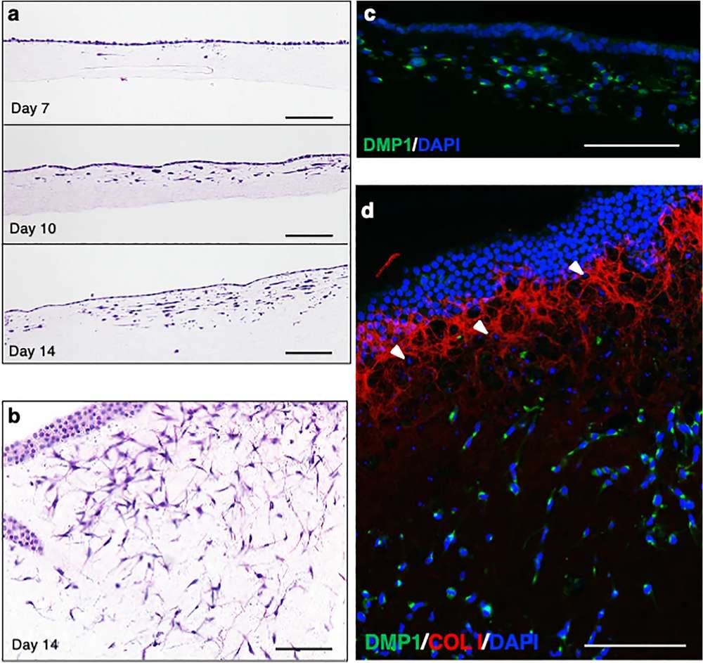 Fig. 1 Induction of osteoblastic and osteocytic cells from hiPSCs (414C2) on type I collagen gel.1,3
The researchers sought to identify the reference genes best suited for experiments that induce osteogenic differentiation from human induced pluripotent stem cells. They were cultured in an undifferentiated maintenance medium and after confluence, further cultured in an osteogenic differentiation medium for 28 days. RT-qPCR was performed on undifferentiation markers, osteoblast and osteocyte differentiation markers, and reference gene candidates. The expression stability of each reference gene candidate was ranked using four algorithms.
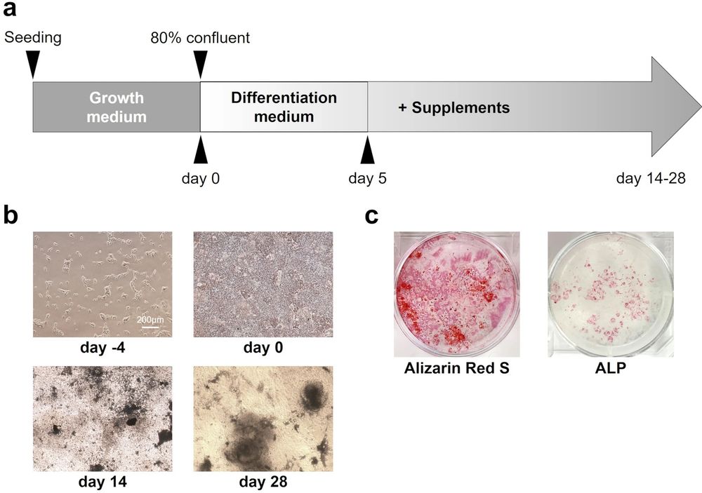 Fig. 2 Osteogenic differentiation in human induced pluripotent stem (iPS) cells.2,3
What Our Clients Say
"Creative Biolabs helped us establish an iPSC-derived osteogenic model for a rare skeletal disease project. The team carefully optimized the differentiation conditions and provided clear marker analysis and mineralization data. Their technical feedback was very helpful for interpreting the phenotype."
— Principal Investigator, Academic Research Institute
"We needed osteogenic cells generated from several patient-specific iPSC lines in parallel. Creative Biolabs designed a workflow that minimized batch variation and provided regular updates throughout the project. The final report was well organized and easy to integrate into our internal study package."
— Senior Scientist, Biotechnology Company
"Our project required osteogenic cells compatible with scaffold-based culture. Creative Biolabs supported the cell preparation, scaffold seeding strategy, and downstream staining analysis. The results helped us compare different material formulations with greater confidence."
— R&D Manager, Regenerative Medicine Company
"We were developing a screening assay for compounds affecting osteoblast maturation. Creative Biolabs provided iPSC-derived osteogenic cultures with ALP and mineralization readouts, and their assay recommendations helped us refine the treatment window."
— Translational Research Lead, Pharmaceutical Company
FAQs
Q: What type of iPSC lines can be used for osteogenic differentiation?
A: We can work with healthy donor iPSCs, patient-derived iPSCs, disease-specific iPSCs, gene-edited clones, reporter lines, and isogenic control pairs. If you do not yet have iPSCs, Creative Biolabs can also support upstream reprogramming and characterization.
Q: What is the typical timeline for iPSC-derived osteogenic differentiation?
A: The timeline depends on the starting line, desired endpoint, QC package, and scale. Early osteogenic progenitor generation may require a shorter workflow, while mature mineralization-positive cultures usually require extended induction and characterization. A project-specific timeline will be provided after technical consultation.
Q: Can you generate mature osteoblast-like cells with mineralization capability?
A: Yes. We can design differentiation workflows aimed at osteoblast-like maturation and mineralized matrix deposition. Functional readouts such as ALP activity, Alizarin Red staining, calcium quantification, and mineralized nodule imaging can be included.
Q: Can iPSC-derived osteogenic cells be used for disease modeling?
A: Yes. Patient-derived or gene-edited iPSCs can be differentiated into osteogenic cells to model skeletal disease phenotypes. Matched control lines and isogenic pairs are especially useful for identifying mutation-specific effects.
Q: Can you compare multiple iPSC lines in parallel?
A: Yes. We frequently support parallel differentiation of control, patient, and gene-edited iPSC lines. Parallel processing helps reduce batch effects and strengthens comparative interpretation.
Q: Are cryopreserved iPSC-derived osteogenic cells available?
A: Depending on the differentiation stage and project design, cryopreserved delivery may be possible. Some mature osteogenic cultures are best delivered as assay-ready live cultures or fixed samples. We will recommend the most suitable format based on your endpoint.
Q: Can you develop a fully customized differentiation protocol?
A: Yes. Creative Biolabs provides custom protocol development for challenging cell lines, rare disease models, special culture systems, or non-standard assay endpoints. Feasibility testing can be performed before full-scale production.
Take the Next Step with Creative Biolabs







1. Contact Us
via the Inquiry Form or Email
2. Define Your Needs
Cell Type, Function, Quantity, Modifications
3. Kickstart the Project
Our Expert Team Guiding Every Step
Creative Biolabs is committed to helping researchers access reliable, human-relevant osteogenic cell models derived from iPSCs. Our iPSC-derived osteogenic cell differentiation services are designed for flexibility, scientific rigor, and practical downstream usability. From early osteogenic commitment to mineralization-competent osteoblast-like cultures, we provide customized solutions that support bone biology research, disease modeling, drug discovery, biomaterial evaluation, and regenerative medicine development.
Contact us today to discuss your iPSC-derived osteogenic cell differentiation project.
References
- Kawai, Shunsuke, et al. "3D osteogenic differentiation of human iPSCs reveals the role of TGFβ signal in the transition from progenitors to osteoblasts and osteoblasts to osteocytes." Scientific Reports 13.1 (2023): 1094. https://doi.org/10.1038/s41598-023-27556-w
- Okamura, Kensuke, et al. "RT-qPCR analyses on the osteogenic differentiation from human iPS cells: an investigation of reference genes." Scientific Reports 10.1 (2020): 11748. https://doi.org/10.1038/s41598-020-68752-2
- Distributed under Open Access license CC BY 4.0, without modification.