Creative Biolabs' Morphology Observation Service delivers non-destructive, cost-effective quality control by combining cellular morphology with transcriptomics, as supported by recent research. It captures dynamic biological processes missed by gene expression alone. Leveraging advanced Cell Painting, deep-learning nucleocentric profiling, and high-content imaging, we provide high-dimensional phenotypic insights, identify subtle sub-cellular changes, and ensure iPSC models meet pluripotency and maturity golden standards for reliable drug discovery.
Discover How We Can Help - Request a Consultation
We assess high-quality iPSC colonies by analyzing epithelial-like structures, intercellular spaces, and duct networks to ensure medium transport, and track morphological evolution via high-content imaging to verify pluripotency and culture quality.
Spontaneous or directed differentiation of iPSCs can be identified by the following morphological alterations:
| Evaluation Index | Pluripotent iPSC Morphology | Differentiated iPSC Morphology |
|---|---|---|
| Colony edges | Smooth, clear, and well-defined | Fuzzy, irregular, and blurred |
| Cell density | Tightly packed | Loosely arranged with gaps |
| Nucleus-to-cytoplasm ratio | High | Low |
| Nucleolus | Prominent | Indistinct |
To initiate the service, clients typically provide Starting Materials such as:
At Creative Biolabs, we provide a complete, high-capacity pipeline for morphological characterization tailored to the rigorous demands of modern biotechnology. Our advantages include:
One-stop morphology service from initial cell line validation and maintenance to high-dimensional phenotypic profiling.
Automated confocal and electron microscopy platforms capable of processing thousands of samples with over 100,000 images per study.
Fully flexible assay design, allowing clients to specify organelle targets, staining combinations, and custom deep-learning model parameters.
Use of proprietary "nucleocentric" segmentation to ensure >96% accuracy in dense, complex neural cultures.
Integration of Quality-by-Design (QbD) and Process Analytical Techniques (PAT) to guarantee data reproducibility.
Inclusion of negative and positive morphological controls to verify phenotype specificity across biological replicates.
High-standard quality control tools and bioinformatics pipelines to quantify over 3,000 morphological features per cell.
Experience the Creative Biolabs Advantage - Get a Quote Today
Induced pluripotent stem cells (iPSCs) enable cellular-level analyses of previously intractable cell types and lineages, yet the full diversity of in vitro-derived cells remains incompletely characterized, including some cell types and states absent in vivo. Single-cell RNA sequencing (scRNA-seq) enhances iPSC-derived cell characterization resolution, but gene expression alone fails to fully reflect cellular functions and disease-driving biological processes, highlighting the need for additional phenotypic measurements. Cell Painting (CP), a high-content imaging technique, is used to expand single-cell phenotypic readouts and conduct combined CP-based morphological and scRNA-seq-based gene expression analyses on iPSC-derived cortical neurons.
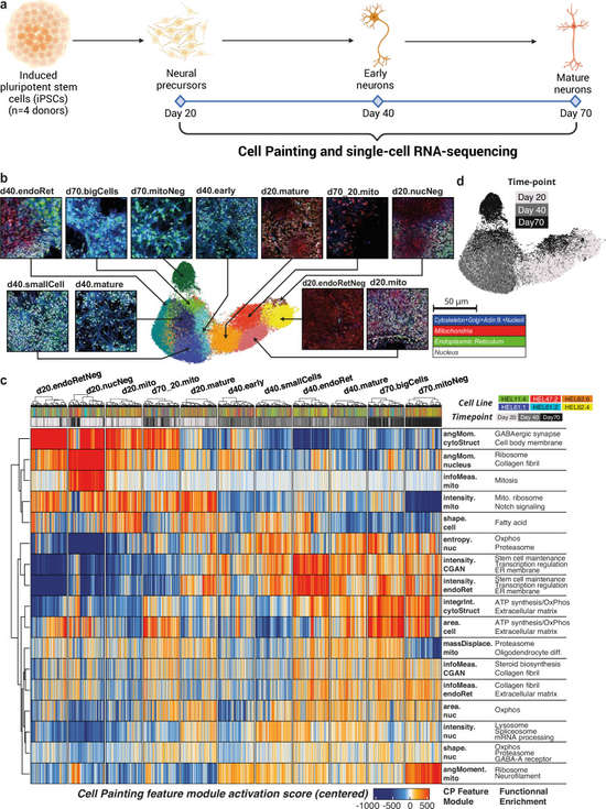 Fig.1 Morphological detection of iPSC differentiation into cortical neurons and scRNA-seq sequencing.1
A: We use a "nucleocentric" deep learning model that focuses on the nuclear ROI and its immediate 18 µm micro-environment. This avoids the common errors associated with overlapping cell membranes in 100% confluent neural cultures.
A: While the standard Cell Painting protocol requires fixation, our non-destructive live-cell imaging alternatives allow for longitudinal tracking of the same culture over time.
A: We benchmark our iPSC-derived morphology data against validated fetal brain references to ensure your in vitro models are physiologically relevant.
A: Yes, we have successfully identified signatures for Schizophrenia, Bipolar Disorder, and various metabolic syndromes by capturing changes in organelle distribution and cell shape.
A: Morphology captures the functional execution of the cell. mRNA levels only partially correlate with protein and function; morphology provides the ground truth of a cell's health and maturity.
Creative Biolabs offers a comprehensive suite of morphology-related services, including high-content Cell Painting, CNN-based cell classification, ultrastructural electron microscopy, and integrated multi-modal data analysis. We provide the biological intelligence needed to de-risk your R&D pipeline.
Contact Our Team for More Information and to Discuss Your Project
Reference
For Research Use Only. Not For Clinical Use.