1. Home
  2. ADC Development
  3. ADC In Vitro Analysis
  4. ADC In Vitro Efficacy Evaluation: Comprehensive Binding, Internalization & Cytotoxicity Assays

ADC In Vitro Efficacy Evaluation: Comprehensive Binding, Internalization & Cytotoxicity Assays

Antibody-Drug Conjugates (ADCs) represent a paradigm shift in targeted cancer therapy, combining the specificity of monoclonal antibodies with the potency of cytotoxic payloads. Creative Biolabs offers a comprehensive ADC in vitro efficacy evaluation solution designed to validate every critical step of the ADC mechanism of action. From antigen binding affinity to payload-induced cytotoxicity, our multi-dimensional assessment platform ensures your candidates possess the optimal biochemical and cellular properties required for preclinical and clinical success. Leveraging advanced analytical platforms and diverse cell-based assays, we provide actionable insights that accelerate the development of next-generation ADC therapeutics.

Request Efficacy Evaluation

Decoding ADC Mechanism of Action through In Vitro Validation

ADC efficacy is determined by a complex cascade of events: target binding, internalization, payload release, and ultimately, cancer cell death. Each step must be rigorously validated in vitro to predict in vivo performance and minimize clinical failure risks. Our integrated evaluation suite provides a comprehensive "mechanistic audit" covering all four pillars of ADC activity.

Core Pillars of Our In Vitro Efficacy Framework

We provide a systematic evaluation matrix for your ADC candidates, focusing on the four most critical parameters for preclinical efficacy assessment:

  • Antigen Binding Affinity: Validating that the conjugation process does not compromise the antibody's ability to recognize and bind to its target antigen with high specificity and avidity.
  • Internalization Efficiency: Quantifying the rate and extent of ADC uptake into target cells, a prerequisite for payload delivery to intracellular compartments.
  • Cytotoxicity Potency: Assessing the direct cell-killing capacity of the ADC, including IC50 determination and apoptosis induction across diverse cancer cell lines.
  • Fc-Mediated Immune Activity: Evaluating the antibody Fc region's contribution to efficacy through immune cell recruitment and activation (ADCC, CDC, ADCP).

Integrated In Vitro Efficacy Evaluation Units

Our service matrix is organized into four intensive evaluation units, each designed to answer fundamental questions about your ADC's functional attributes and mechanism of action.

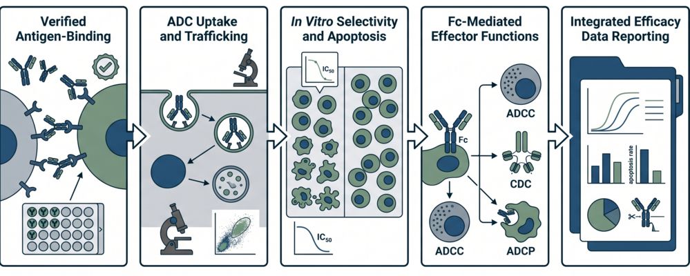
Antigen Binding Affinity Validation

The foundation of ADC efficacy is the retention of antigen-binding capacity after conjugation. This unit verifies that the antibody component maintains high-affinity interaction with its target receptor on cancer cell surfaces.

Critical Challenges Solved
  • Affinity Loss Detection: Identifying conjugation-induced conformational changes that reduce target binding avidity.
  • On-Target Specificity: Confirming selective binding to tumor-associated antigens versus off-target receptors.
  • Binding Kinetics: Measuring association (ka) and dissociation (kd) rates to establish the stability of ADC-target complexes.
Technical Platforms
We utilize Surface Plasmon Resonance (SPR) for real-time kinetic analysis and Flow Cytometry (FACS) for cell-surface binding quantification. ELISA-based assays provide high-throughput screening, while Radioligand Binding Assays offer ultra-sensitive detection limits.

Internalization Efficiency Profiling

Internalization is the rate-limiting step for payload delivery. This unit quantifies the dynamics of ADC uptake, trafficking, and accumulation in intracellular compartments.

Critical Challenges Solved
  • Uptake Kinetics: Determining the time-dependent internalization rate (T1/2) to optimize dosing schedules.
  • Trafficking Validation: Confirming delivery to lysosomal compartments where payload release occurs.
  • Recycling vs. Degradation: Differentiating between ADCs that are degraded for payload release versus those recycled back to the cell surface.
Technical Platforms
We employ Fluorescent Microscopy for spatiotemporal tracking and Flow Cytometry for quantitative uptake analysis. Radiolabeled ADCs provide precise internalization quantitation, while Confocal Laser Scanning Microscopy (CLSM) visualizes subcellular localization.

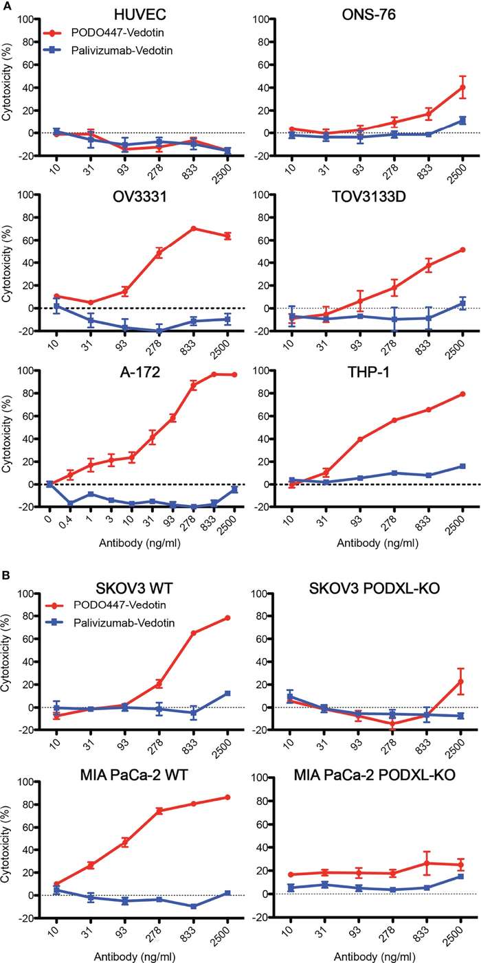
In Vitro Cytotoxicity Potency Assessment

The ultimate measure of ADC success is the ability to induce cancer cell death. This unit evaluates the direct cytotoxic impact of the conjugated payload across diverse cell line models.

Critical Challenges Solved
  • IC50 Determination: Establishing dose-response curves to define the concentration required for 50% cell death.
  • Selectivity Index: Comparing potency in target-positive versus target-negative cell lines to confirm on-target killing.
  • Mechanism of Cell Death: Distinguishing apoptosis, necrosis, and cell-cycle arrest through multiparametric assays.
Technical Platforms
We utilize MTT/MTS/XTT Assays for cell viability, Annexin V/Propidium Iodide Staining for apoptosis detection, and Cell-Cycle Analysis (PI Staining) for proliferation profiling. Real-Time Cell Analysis (RTCA) provides label-free, kinetic cytotoxicity monitoring.

Fc-Mediated Immune Cytotoxicity Evaluation

Beyond direct payload toxicity, the antibody Fc region can engage immune effector cells. This unit assesses the "secondary efficacy" contributed by Fc-mediated mechanisms.

Critical Challenges Solved
  • Effector Cell Recruitment: Quantifying the ability of ADCs to recruit NK cells, macrophages, and complement proteins.
  • Fc Gamma Receptor (FcγR) Binding: Profiling interactions with activating versus inhibitory FcγRs to predict clinical efficacy.
  • Synergistic Killing: Evaluating the combined effect of payload cytotoxicity and immune-mediated cell death.
Technical Platforms
We employ Antibody-Dependent Cellular Cytotoxicity (ADCC) Assays using primary NK cells, Complement-Dependent Cytotoxicity (CDC) Assays with human serum, and Antibody-Dependent Cellular Phagocytosis (ADCP) Assays using monocyte-derived macrophages. FcγR Binding Arrays provide comprehensive Fc effector profiling.

Standardized Workflow for ADC In Vitro Efficacy Evaluation

Our systematic preclinical workflow ensures comprehensive mechanistic validation from target engagement to cell death:

Integrated workflow for ADC in vitro efficacy evaluation

Step 1: Target Binding Affinity Assessment

Verification of antigen-binding capacity using SPR, FACS, or ELISA to confirm that conjugation does not compromise target recognition and avidity.

Step 2: Internalization Kinetics Profiling

Quantitative assessment of ADC uptake, trafficking, and lysosomal delivery using flow cytometry, microscopy, or radiolabel tracing.

Step 3: In Vitro Cytotoxicity Evaluation

Comprehensive cell viability, apoptosis, and cell-cycle analysis across target-positive and target-negative cell lines to establish selectivity and potency.

Step 4: Fc-Mediated Effector Function Analysis

Evaluation of ADCC, CDC, and ADCP activities to quantify the antibody Fc region's contribution to the overall efficacy profile.

Step 5: Integrated Efficacy Data Reporting

Comprehensive data package compilation with IC50 values, mechanism of action summaries, and preclinical efficacy predictions for in vivo study design.

Analytical Platforms for In Vitro Efficacy Assessment

Our integrated evaluation platform combines advanced analytical instrumentation with validated assay protocols to deliver precise, reproducible efficacy data for your ADC candidates.

Surface Plasmon Resonance (SPR)

Real-time kinetic analysis for binding affinity measurement with sub-nanomolar sensitivity.

Flow Cytometry (FACS)

High-throughput cell surface binding and internalization quantification across diverse cell panels.

Confocal Microscopy

Subcellular localization imaging to track ADC trafficking and lysosomal payload delivery.

Real-Time Cell Analysis (RTCA)

Label-free kinetic cytotoxicity monitoring for continuous cell viability profiling.

Multi-Parameter Cytometry

Apoptosis and cell-cycle analysis using Annexin V/PI and propidium iodide staining.

Immune Effector Assays

ADCC, CDC, and ADCP evaluation using primary immune cells and complement systems.

Research Insights: Validating Tumor-Specific Efficacy through Integrated Assays

According to the study by Canals Hernaez et al. (2022), the development of highly specific ADCs like PODO447-Vedotin relies on rigorous in vitro validation to ensure tumor-restricted activity while sparing normal tissues. Their research on Podocalyxin (Podxl) demonstrates how multi-parameter efficacy profiling de-risks clinical development:

Key Findings from the PODO447-ADC Study:

  • Glycan-Specific Binding: Cell-based glycan arrays confirmed that PODO447 targets a tumor-restricted core 1 O-glycan on the Podxl backbone, providing exquisite specificity for malignant cells over normal vascular endothelia.
  • Efficient Internalization: Time-lapse monitoring using dyes conjugates demonstrated robust internalization of the ADC into acidic intracellular compartments of ovarian and pancreatic cancer cells.
  • Potent Cytotoxicity: In vitro MTT assays established sub-nanomolar to low nanomolar IC50 values (e.g., 31 ng/ml in THP-1 and 93 ng/ml in A-172), highlighting the ADC's high therapeutic potency across diverse solid and liquid tumor models.
  • Proven Safety Index: The ADC showed negligible toxicity against normal endothelia (HUVEC), validating its tumor-specific mechanism and predicting a favorable safety profile for in vivo applications.

This integrated efficacy framework enables the identification of candidates with optimal tumor targeting and intracellular delivery dynamics.

In vitro cytotoxicity and selectivity analysis of PODO447-Vedotin.

Fig.1 In vitro cytotoxicity and selectivity of PODO447-Vedotin.1,2

FAQs: ADC In Vitro Efficacy Evaluation

Q: How do you ensure that conjugation does not compromise antigen-binding affinity?

A: We perform side-by-side affinity comparisons between the unconjugated antibody and the ADC using SPR and FACS. This allows us to quantify any conjugation-induced affinity loss and optimize conjugation conditions to preserve target binding.

Q: What cell line models do you recommend for in vitro cytotoxicity evaluation?

A: We recommend a panel of cell lines spanning a range of target antigen expression levels (high, medium, low, and negative) to establish the therapeutic window. This panel should include both the intended indication models and potential off-target toxicity models.

Q: Can you differentiate between direct payload toxicity and Fc-mediated killing?

A: Yes. We use Fc receptor-blocking antibodies or FcgR knockout cell lines to isolate direct cytotoxicity from Fc-mediated effects. Additionally, we perform ADCC/CDC assays with appropriate effect or cells to quantify the Fc contribution.

Q: How do you assess the bystander killing effect in vitro?

A: We employ co-culture systems mixing target-antigen-positive and target-antigen-negative cells at various ratios. The killing of antigen-negative cells indicates bystander effect, which is particularly important for ADCs with membrane-permeable payloads like MMAE or Dxd.

Q: Do you provide 3D cell culture models for efficacy evaluation?

A: Yes. We offer spheroid and organoid models that better mimic the tumor microenvironment and drug penetration barriers. 3D efficacy data often correlates better with in vivo outcomes and can reveal penetration limitations not apparent in 2D monolayer assays.

References:
1. Canals Hernaez, Diana, et al. "Targeting a tumor-specific epitope on podocalyxin increases survival in human tumor preclinical models." Frontiers in Oncology 12 (2022): 856424.https://doi.org/10.3389/fonc.2022.856424
2. Distributed under Open Access License CC BY 4.0, without modification.

For Research Use Only. NOT FOR CLINICAL USE.


Online Inquiry

Name:
Phone:
*E-mail Address:
*Products or Services Interested:
Company/Institution
Project Description:

Welcome! For price inquiries, please feel free to contact us through the form on the left side. We will get back to you as soon as possible.