1. Home
  2. ADC Development
  3. ADC In Vitro Analysis
  4. 3D Cell Culture for ADC Evaluation: Targeting, Penetration & Efficacy Assays

3D Cell Culture for ADC Evaluation: Targeting, Penetration & Efficacy Assays

As an industrial leader in therapeutic antibody development, complex organic synthesis, and bio-conjugates preparation, Creative Biolabs has been actively exploring innovative technologies to advance targeted immunotherapies. Using our advanced 3D cell culture platform, we provide comprehensive 3D cell culture-based in vitro evaluation services for characterizing your candidate drugs using three-dimensional cell clusters that mimic solid tumor architecture. Our services bridge the gap between 2D monolayer assays and in vivo studies, providing more accurate ADC function profiles for solid tumor therapeutics.

Request 3D Evaluation Services

Revolutionizing ADC Evaluation with 3D Cell Culture Technology

Conventional 2D cell cultures, with their flat-bottom matrix and single-cell layer architecture, are insufficient for evaluating Antibody-Drug Conjugates (ADCs) targeting solid tumors. Our 3D cell culture platform bridges the critical gap between simplistic 2D monolayer assays and complex in vivo studies, providing physiologically relevant models that mimic the architectural complexity of solid tumors.

Core Advantages of 3D Cell Culture Models

We provide a comprehensive evaluation matrix leveraging 3D cell culture technology to address the fundamental limitations of traditional 2D assays:

  • Architectural Fidelity: 3D cell clusters replicate the multi-layer structure, extracellular matrix composition, and cell-cell interactions of solid tumors.
  • Penetration Barrier Modeling: The 3D architecture creates diffusion limitations and heterogeneous antigen expression, critical for evaluating ADC tumor penetration efficiency.
  • Microenvironment Complexity: 3D models incorporate hypoxia gradients, pH variations, and stromal cell interactions that influence ADC efficacy.
  • In Vitro-In Vivo Correlation: 3D culture data shows superior correlation with in vivo xenograft outcomes compared to 2D monolayer assays.

Integrated 3D Evaluation Service Units

Our service matrix is organized into five comprehensive evaluation units, each designed to characterize different aspects of ADC performance in 3D cell culture models.

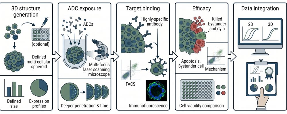
ADC Targeting Profile in 3D Models

Evaluates the ability of ADCs to specifically bind to target antigens in 3D cell clusters, accounting for penetration barriers and heterogeneous antigen expression.

Critical Challenges Solved
  • Penetration-Dependent Binding: Quantifying target binding in outer vs. inner cell layers of 3D spheroids.
  • Heterogeneous Antigen Expression: Mapping antigen density variations across 3D architecture.
  • Affinity vs. Accessibility: Differentiating between affinity limitations and physical penetration barriers.
Technical Platforms
We utilize Confocal Laser Scanning Microscopy (CLSM) for Z-stack imaging of ADC binding, Flow Cytometry on dissociated 3D cells, and Quantitative Immunofluorescence for spatial binding profiling.

ADC Tumor Penetration Assessment

Quantifies the depth and efficiency of ADC penetration into 3D cell clusters, a critical parameter for solid tumor efficacy.

Critical Challenges Solved
  • Penetration Depth Quantification: Measuring how deeply ADCs penetrate into 3D spheroids using fluorescence imaging.
  • Time-Dependent Penetration: Establishing penetration kinetics and equilibrium distribution.
  • Size and Charge Effects: Evaluating how ADC biophysical properties influence penetration efficiency.
Technical Platforms
We employ Confocal Microscopy with Z-stack Analysis, Multiphoton Microscopy for deep-tissue imaging, and Quantitative Fluorescence Analysis using plate readers or image analysis software.

In Vitro Efficacy Evaluation in 3D

Assesses ADC cytotoxicity and cell-killing efficacy in 3D cell culture models that better mimic in vivo solid tumor responses.

Critical Challenges Solved
  • 3D Cytotoxicity Profiling: Establishing IC50 values in spheroids vs. monolayers to quantify the 3D efficacy gap.
  • Bystander Effect in 3D: Evaluating payload diffusion and bystander killing in heterogeneous 3D models.
  • Viability Gradient Analysis: Mapping cell death patterns from outer proliferating layers to inner necrotic cores.
Technical Platforms
We utilize MTT/MTS Assays in 3D, ATP-Based Luminescence Assays, Live/Dead Staining with Confocal Imaging, and Apoptosis Detection (Caspase 3/7 activity).

2D vs 3D Comparative Analysis

Provides side-by-side comparison of ADC performance in 2D monolayer vs. 3D cell culture models to highlight the added value of 3D evaluation.

Critical Challenges Solved
  • Efficacy Correlation: Quantifying the correlation between 2D IC50 and 3D IC50 to establish translation factors.
  • False-Positive Identification: Identifying ADC candidates that show potency in 2D but fail in 3D models.
  • Model Selection Guidance: Providing data-driven recommendations for when to use 2D vs. 3D models in lead optimization.
Technical Platforms
We perform Parallel 2D/3D Cytotoxicity Assays, Comparative Flow Cytometry (apoptosis, cell-cycle), and Bioinformatics Analysis to establish predictive models for in vivo efficacy.

Solid Tumor Efficacy in 3D Models

Evaluates ADC efficacy specifically in solid tumor models, incorporating stromal cells and extracellular matrix components to enhance physiological relevance.

Critical Challenges Solved
  • Stromal Barrier Effects: Assessing how cancer-associated fibroblasts and matrix proteins impede ADC access.
  • Hypoxia-Induced Resistance: Evaluating ADC efficacy under hypoxic conditions that mimic tumor microenvironments.
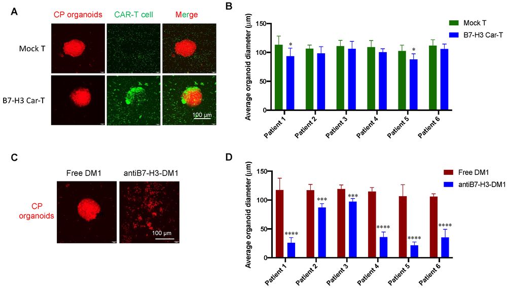
  • Patient-Derived Model Validation: Testing ADCs in patient-derived organoids or spheroids for personalized medicine applications.
Technical Platforms
We employ Co-culture Spheroid Models (cancer cells + stromal cells), Hypoxia Chamber Cultures, Patient-Derived Organoid (PDO) Assays, and Extracellular Matrix (ECM) Embedded Cultures.

Standardized Workflow for 3D Cell Culture Evaluation

Our systematic workflow ensures reproducible and physiologically relevant evaluation of ADC candidates using 3D cell culture models:

Integrated workflow for 3D cell culture-based ADC evaluation

Step 1: 3D Model Establishment

Culture and characterization of 3D cell clusters (spheroids, organoids) with defined size, architecture, and antigen expression profiles.

Step 2: ADC Exposure and Penetration

Controlled ADC treatment with time-dependent penetration tracking using confocal microscopy and quantitative fluorescence analysis.

Step 3: Targeting and Binding Assessment

Evaluation of ADC binding profiles in 3D architecture using immunofluorescence, flow cytometry, and spatial analysis.

Step 4: Efficacy and Cytotoxicity Analysis

Comprehensive cell viability, apoptosis, and bystander effect assessment in 3D models using multi-parameter assays.

Step 5: Data Integration and Reporting

Compilation of 3D evaluation data with 2D comparative analysis, providing actionable insights for ADC optimization and in vivo study design.

Advanced Platforms for 3D Cell Culture Evaluation

Our integrated 3D evaluation platform combines advanced cell culture technologies with sophisticated analytical instrumentation to deliver physiologically relevant ADC characterization data.

3D Spheroid Culture

Uniform spheroid generation using ultra-low attachment plates or hanging drop methods.

Organoid Culture

Patient-derived organoid models preserving native tumor heterogeneity and drug response.

Confocal Microscopy

High-resolution Z-stack imaging for penetration depth and binding distribution analysis.

Multiphoton Microscopy

Deep-tissue imaging for thick 3D models with minimal photodamage.

3D Cytotoxicity Assays

ATP-based luminescence and MTT/MTS assays optimized for 3D cell clusters.

Bioinformatics Analysis

Data integration and predictive modeling for in vitro-in vivo correlation establishment.

Research Insights: Advancing B7-H3 Targeted ADC Evaluation in 3D Organoid Models

Recent research by Tang et al. (2022) has demonstrated the critical importance of using 3D organoid models for evaluating B7-H3 targeted immunotherapies, particularly in rare epithelial tumors like Craniopharyngioma (CP). This study provides a paradigm shift in how we assess the penetration and efficacy of Antibody-Drug Conjugates (ADCs) versus other modalities like CAR-T cells.

Key Research Highlights:

  • First CP Organoid Establishment: The researchers established the first in vitro 3D organoid model of CP using fresh clinical specimens, preserving the histological characteristics and marker expression (B7-H3, CD133, CK-7, CTNNB1) of native tumor tissues.
  • High B7-H3 Expression Profiling: B7-H3 was identified as a highly and homogeneously expressed pan-cancer antigen in CP, showing the highest expression levels among twelve different cancer types evaluated.
  • 2D vs. 3D Efficacy Discrepancy: While B7-H3 targeted CAR-T cells showed potent killing in 2D models, their efficacy was significantly limited in 3D organoids, likely due to restricted T-cell trafficking and infiltration throughout the complex 3D architecture.
  • ADC Superiority in Solid Tumors: In contrast to CAR-T, the B7-H3-targeted antibody-DM1 conjugate exhibited potent tumor suppression in both 2D and 3D models, confirming its robust penetration and superior efficacy profile for solid tumor therapeutics.

The CP organoid model serves as a superior platform for drug screening, revealing that ADC-based strategies may overcome the physical barriers that impede cellular therapies in solid epithelial tumors.

Antitumor effects of B7-H3-targeted CAR-T cells and ADCs in CP organoid model.

Fig.1 Antitumor activity evaluation of B7-H3-targeted CAR-T cells and ADCs in CP organoid models.1,2

FAQs: 3D Cell Culture based In Vitro Evaluation

Q: Why is 3D cell culture superior to 2D monolayer for ADC evaluation?

A: 3D cell cultures mimic the architectural complexity, cell-cell interactions, penetration barriers, and microenvironmental factors of solid tumors. This physiological relevance provides more accurate prediction of in vivo efficacy compared to 2D monolayers, which lack these critical features.

Q: What types of 3D models do you recommend for ADC evaluation?

A: We recommend a combination of spheroid models (for penetration and efficacy screening), organoid models (for patient-specific response prediction), and co-culture models (for stromal barrier evaluation). The specific model selection depends on the research question and therapeutic context.

Q: How do you quantify ADC penetration in 3D spheroids?

A: We use confocal microscopy with Z-stack imaging to visualize and quantify ADC distribution across spheroid layers. Quantitative fluorescence analysis measures penetration depth, binding heterogeneity, and time-dependent distribution profiles.

Q: Can you establish in vitro-in vivo correlation using 3D models?

A: Yes. We perform parallel evaluation of ADC candidates in 3D models and subsequent in vivo xenograft studies to establish correlation models. This data enables prediction of in vivo efficacy based on 3D in vitro screening results.

Q: Do you provide patient-derived organoid models for personalized ADC evaluation?

A: Yes. We offer patient-derived organoid (PDO) models for evaluating ADC efficacy in patient-specific contexts. This service is particularly valuable for personalized medicine applications and clinical trial design.

References:
1. Tang, Mei, et al. "Evaluation of B7-H3 targeted immunotherapy in a 3D organoid model of craniopharyngioma." Biomolecules 12.12 (2022): 1744. https://doi.org/10.3390/biom12121744
2. Distributed under Open Access License CC BY 4.0, without modification.

For Research Use Only. NOT FOR CLINICAL USE.


Online Inquiry

Name:
Phone:
*E-mail Address:
*Products or Services Interested:
Company/Institution
Project Description:

Welcome! For price inquiries, please feel free to contact us through the form on the left side. We will get back to you as soon as possible.