- Home
- ADC Development
- ADC In Vitro Analysis
- 3D Cell Culture for ADC Evaluation: Targeting, Penetration & Efficacy Assays
- ADC Tumor Penetration Analysis: 3D Spheroid Models for Tissue Distribution
ADC Tumor Penetration Analysis: 3D Spheroid Models for Pre-clinical Tissue Distribution
Deep tissue penetration is a critical parameter for antibody-drug conjugates (ADCs) to achieve clinical efficacy in solid tumors. Creative Biolabs provides comprehensive tumor penetration analysis services using advanced 3D spheroid models. Our static and fluidic penetration platforms help characterize the tissue distribution, diffusion kinetics, and penetration depth of your antibody-drug conjugate (ADC) candidates—delivering critical data to support pre-clinical lead optimization.
Inquire About Penetration EvaluationOverview: The Challenge of Solid Tumor Penetration in ADC Therapeutics
Unlike hematological malignancies, solid tumors present massive physical and physiological barriers that severely limit macromolecular drug transport. Dense extracellular matrix (ECM), tight cell-cell junctions, and high interstitial fluid pressure (IFP) combine to restrict passive diffusion, often trapping large antibody-based therapeutics near the blood vessels at the tumor periphery.
Why Tissue Penetration is Critical for ADC Pre-Clinical Success
In solid tumor indications, the spatial distribution of an ADC directly dictates its therapeutic outcome. Key considerations include:
- • The Binding Site Barrier (BSB) Effect: Paradoxically, ADCs with extremely high binding affinities can bind rapidly to target receptors encountered on the outermost cells of a tumor mass, exhausting the drug supply and preventing deep diffusion.
- • Eliminating Antigen-Low Tumor Cores: Dense solid tumors often exhibit target antigen heterogeneity. If the ADC cannot diffuse deeply to reach cells in the inner necrotic or hypoxic core, these low-expressing cells will survive and drive tumor recurrence.
- • Bystander Effect Maximization: For ADCs leveraging membrane-permeable payloads, understanding the exact transport and distribution of released toxin molecules through neighboring tissue is key to maximizing bystander killing.
Physiologically Relevant 3D In Vitro Evaluation
Our pre-clinical tumor penetration analysis bypasses the limitations of standard 2D culture. By establishing tumor-stroma co-culture spheroids, patient-derived organoids, and microfluidic platforms, we accurately replicate the physical barriers and fluid dynamics of solid tumors—delivering precise predictions of in vivo transport behavior.
Overcoming Key Hurdles in Tissue Transport Characterization
Measuring the physical transport of macromolecules within dense biological structures presents unique analytical difficulties that require specialized technology:
- ▶ Loss of Native Barriers in 2D: Cells grown on flat plastic lack extracellular matrices and tight cell junctions, meaning they cannot model resistance to macromolecular diffusion.
- ▶ Imaging Depth Limits: Standard fluorescence microscopy cannot penetrate deep into opaque 3D spheroids, requiring advanced optical sectioning and chemical tissue clearing.
- ▶ Passive vs. Active Transport: Modeling transport under purely static conditions fails to account for microvascular flow, extravasation, and mechanical shear stress experienced in vivo.
Our 3D Tumor Penetration Analysis Services
We provide comprehensive penetration analysis services designed to quantify transport mechanics, spatial distribution, and core accumulation of ADCs in advanced tissue models:
Tailored Penetration Solutions for Your Program
Each solid tumor indication presents unique extracellular and physical transport barriers. Our platforms can be fully customized to use tumor-specific cell lines, patient-derived material, or specialized microfluidic chips. Whether you need rapid screening of antibody fragments or highly detailed, high-resolution 3D cleared tissue imaging under active flow, our scientists will design the optimal preclinical evaluation scheme.
| Service Name | Technical Specifications | Analysis Capabilities | Service Deliverables |
|---|---|---|---|
|
Primary Method Static 3D Spheroid Diffusion Assay Spatiotemporal monitoring of fluorophore-tagged ADC penetration in standardized tumor spheroids. |
• Models: Monotypic or heterotypic (tumor + fibroblast) 3D spheroids (MCTS). • Imaging: Automated laser scanning confocal microscopy. • Time-course: 1h, 4h, 8h, 24h, up to 48h incubation. • Analysis: Concentric ring pixel intensity profiling. |
• Radial diffusion velocity calculation • Max penetration depth profiling • Relative accumulation comparison • Core vs. peripheral density ratio |
• Confocal optical slice image gallery • Time-dependent radial profile charts • Comparative transport report • Processing and QC records |
|
Fluidic Flow Microfluidic Perfused Penetration Service Perfused tumor spheroid analysis simulating capillary flow, shear stress, and active extravasation. |
• System: Organ-on-a-chip or microfluidic flow cell systems. • Barriers: Endothelial cell monolayer interface on collagen matrices. • Flow Rate: Controlled shear stress (0.1–1.0 dyn/cm²). • Spheroids: Hydrogel-embedded tumor micro-tissues. |
• Trans-endothelial extravasation kinetics • Transport under continuous shear flow • Vascular clearance vs. retention profiling • Dynamic transport index generation |
• Real-time video of endothelial crossing • Volumetric penetration profile over time • Dynamic transport summary table • Specialized microfluidic data package |
|
Ultra-Resolution Cleared Tissue Volumetric Profiling Advanced tissue clearing coupled with light-sheet microscopy for deep, high-resolution 3D transport mapping. |
• Clearing Method: Hydrogel-based clearing (CLARITY) or solvent clearing (BABB/iDISCO). • Instrumentation: High-resolution Light-Sheet Fluorescence Microscopy (LSFM). • Spheroid Size: Large spheroids (>500 μm) or fresh tumor organoids. • Resolution: Subcellular resolution in full 3D volumes. |
• True 3D volumetric transport mapping • Deep-core fluorophore density quantification • ECM collagen/fibronectin co-localization • Cellular binding site barrier evaluation |
• 3D volumetric cleared-tissue video • Spatial distribution intensity curves • Co-localization analysis reports • Cleared-sample preparation logs |
|
Comparative Affinity vs. Penetration Profiling Direct assessment of how antibody affinity and target receptor expression alter tissue penetration. |
• CANDIDATES: Multiple ADC clones or variants with graded affinities. • Expression: Cell lines with high, medium, and low antigen density. • Analytical Run: Parallel high-throughput confocal z-profiling. • Target: Identification of the "affinity threshold" for deep core transport. |
• Quantifying the Binding Site Barrier effect • Correlation: Affinity vs. Penetration Velocity • Optimal affinity window mapping • Target-density dependent transport kinetics |
• Binding Site Barrier evaluation charts • Candidate ranking and recommendation report • Affinity-penetration correlation graphs • Expert pre-clinical consultation |
Specialized Preclinical Evaluations
ECM Modification Profiling
Evaluation of penetration velocity and depth in spheroids pre-treated with ECM-remodeling agents (e.g., hyaluronidase or collagenase), providing rationale for combination therapies to enhance ADC transport.
Patient-Derived Organoid Homing
Evaluating penetration in patient-derived cancer organoids (PDOs), conserving tumor phenotypic diversity and microarchitectures for highly translatable preclinical candidate evaluation.
Trans-tissue Bystander Modeling
Tracing the spatial migration and killing efficiency of bystander payloads across multiple layers of non-target cells inside co-culture spheroids to guide linker-payload selection.
IND Submission-Ready Packages
Fully documented 3D transport validation packages, system suitability records, and quantitative depth analysis datasets, structured according to preclinical documentation standards.
Standardized Workflow for 3D Tumor Penetration Analysis
We apply a strict, quality-controlled multi-phase process to guarantee accurate, reproducible tissue transport profiles:
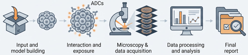
Phase 1: Labeled Conjugation & Integrity Control
ADC candidates are conjugated with stable, bright fluorophores using optimized chemistry. Strict QC checks, including Size Exclusion Chromatography (SEC) and UV/Vis, guarantee conjugation does not alter antigen-binding affinity or prompt aggregation.
Phase 2: Uniform 3D Spheroid/Organoid Culture
Spheroids are generated using low-attachment plates (ULA) with co-cultures of tumor cells and extracellular matrix-producing fibroblasts. Cultures are grown to a uniform size (typically 400-500 μm) to replicate native physical transport resistance.
Phase 3: Kinetic Incubation & Spatiotemporal Imaging
Labeled ADCs are incubated with the live spheroids under static or dynamic microfluidic flow conditions. Concentric optical slicing is performed via multi-laser confocal microscopy at pre-defined time intervals (up to 48 hours).
Phase 4: Optical Tissue Clearing & LSFM Volumetric Run
For deep, high-resolution mapping, samples are optically cleared using validated chemical clearing methods (CLARITY or solvent-based clearing). Cleared tissues are analyzed via Light-Sheet Fluorescence Microscopy (LSFM) for seamless 3D volumetric mapping.
Phase 5: Digital Transport Modeling & Reporting
Advanced digital image processing is used to calculate radial distribution profiles, core-to-periphery ratios, and diffusion coefficients. We deliver a comprehensive pre-clinical candidate transport ranking report.
Advanced Platforms for 3D Tumor Penetration Analysis
We combine advanced 3D cell culture systems with state-of-the-art optical sectioning and microfluidic technologies to characterize macromolecular transport with extreme precision:
1. Static 3D Spheroid Diffusion Platform
Our baseline platform for high-throughput screening of transport characteristics. Standardized monotypic or heterotypic tumor spheroids provide uniform extracellular matrix and cell-junction barriers to evaluate passive transport mechanics.
- • High-throughput Screening: Standardized ULA 96-well format for rapid parallel comparison of multiple candidates.
- • Uniform Size Control: Spheroids are strictly qualified to 400–500 μm diameter with <5% variation.
- • Direct Transport Comparisons: Side-by-side screening of varying molecular formats (mAbs, Fabs, ADCs).
2. Microfluidic Shear & Perfused Platform
A cutting-edge microfluidic organ-on-a-chip platform that integrates endothelial barriers with 3D tumor matrices. Simulates capillary blood flow, endothelial extravasation, and interstitial fluid pressure.
- • Shear Stress Control: Replicates microvascular shear dynamics (0.1–1.0 dyn/cm²) using digital flow control pumps.
- • Extravasation Profiling: Measures active extravasation across qualified HUVEC endothelial monolayers.
- • Dynamic Transport index: Evaluates transport kinetics under continuous replenishment conditions.
3. Multichannel Laser Scanning Confocal Platform
High-resolution confocal laser scanning microscopy (CLSM) equipped with multiple laser lines to resolve fluorescently labeled ADCs, target receptors, and organelle markers in the outer 100-200 μm layers of intact spheroids.
- • Spectral Unmixing: Resolves complex multi-fluorophore combinations without spectral bleed-through.
- • Time-Lapse Live Imaging: Environmentally controlled stage incubators track penetration in live cells in real-time.
- • Radial Distribution Profiling: Automated image processing algorithms profile pixel-intensity distribution in concentric shells.
4. Cleared Tissue & Light-Sheet Volumetric Platform
Designed for full-volume 3D mapping of large spheroids and patient-derived organoids. Optical tissue clearing eliminates light scattering, enabling light-sheet microscopy to map subcellular transport depths in three dimensions.
- • Isotropic Resolution: Light-Sheet Fluorescence Microscopy (LSFM) provides high z-axis resolution across deep tissue structures.
- • Total Sample Imaging: Maps transport depths up to 1000 μm without optical fading.
- • 3D Reconstruction: Digital volumetric rendering software maps spatial accumulation in 3D space.
Why Choose Our 3D Tumor Penetration Analysis Services?
Physiologically Relevant 3D Models
We use monotypic/heterotypic spheroids, organoids, and tissue slices that preserve extracellular barriers, cell-cell junctions, and transport resistances for realistic predictions.
High-Resolution Confocal & Cleared Imaging
Our advanced confocal and tissue clearing platforms provide high-resolution, deep-tissue imaging for volumetric reconstruction and quantitative depth mapping.
Affinity-Penetration Correlation Analysis
We analyze the physical relationship between antibody affinity, target receptor expression density, and tissue penetration depth to determine the optimal affinity window.
Accelerated Turnaround for Pre-clinical Programs
Our standardized workflow and dedicated imaging team deliver comprehensive targeting and penetration analysis reports within 3-4 weeks to keep your timeline on track.
Research Insights: Overcoming the Binding Site Barrier to Improve Solid Tumor Accumulation
Recent advances in macromolecular transport kinetics have confirmed that extremely high antibody-antigen binding affinity can create a "binding site barrier" that limits solid tumor penetration. According to Canals Hernaez et al. (2022), ADCs designed with optimized moderate binding affinities or those targeting tumor-specific glyco-epitopes often exhibit far more uniform spatial distribution and superior therapeutic indices compared to very high-affinity counterparts.
Key Insights from Recent Pre-clinical Research:
- • The Binding Site Barrier: When binding affinity is excessively high, ADC molecules bind rapidly to target cells located near vascular exits, trapping the macromolecule at the tumor periphery and leaving the inner tumor core untreated.
- • Affinity Tuning: Reducing binding affinity slightly (e.g., from sub-nanomolar to low nanomolar KD) can paradoxically improve tumor accumulation and therapeutic index by allowing deeper, more homogeneous tissue penetration.
- • Microenvironment remodeling: Pre-treating 3D models with matrix-remodeling enzymes or vascular-permeabilizing agents is shown to significantly enhance penetration velocity and core accumulation of ADCs (Matsuda et al., 2020).
These findings emphasize the importance of analyzing tissue transport kinetics—rather than relying solely on 2D affinity measurements—during early candidate characterization.

Fig.1 Time-course z-stack confocal imaging maps the spatial transport of labeled ADC candidates through a 3D tumor spheroid.1,3
FAQs about ADC Tumor Penetration Analysis
Q: Why is tumor penetration critical for solid tumor ADC therapeutics?
A: Solid tumors present physical barriers like dense ECM and high interstitial fluid pressure that limit antibody diffusion. Evaluating penetration ensures ADCs can reach cells deep within the tumor core, preventing survival of antigen-low or negative cells.
Q: What is the binding site barrier, and how does it affect ADC distribution?
A: The binding site barrier occurs when an ADC binds too tightly to target receptors at the tumor periphery, preventing it from diffusing deeper. Our assays help evaluate if tuning affinity down can enhance overall penetration and tumor accumulation.
Q: How do 3D tumor spheroids model in vivo tissue penetration better than 2D cultures?
A: 3D spheroids develop realistic cell junctions, extracellular matrix, and nutrient/oxygen gradients. This mimics solid tumor architecture and physical transport barriers, providing predictive data for in vivo translation.
Q: What is the difference between static and fluidic 3D penetration models?
A: Static models evaluate passive diffusion through spheroids. Fluidic models (using microfluidics) introduce active flow and shear stress, simulating vascular transport and extravasation alongside tissue diffusion.
Q: How much ADC sample is required for a complete 3D penetration study?
A: We typically require 100-200 μg of purified ADC for standard confocal profiling. If tissue clearing or multiple timepoints are needed, 300-500 μg is recommended.
Other Pre-clinical Analysis Solutions
Related Products
Related Resources

References:
1. Canals Hernaez, Diana, et al. "Targeting a tumor-specific epitope on podocalyxin increases survival in human tumor preclinical models." Frontiers in Oncology 12 (2022): 856424. https://doi.org/10.3389/fonc.2022.856424
2. Matsuda, Y., Leung, M., Okuzumi, T., Mendelsohn, B. A Purification Strategy Utilizing Hydrophobic Interaction Chromatography to Obtain Homogeneous Species from a Site-Specific Antibody Drug Conjugate. Antibodies (Basel). 2020;9(2):16. https://doi.org/10.3390/antib9020016
3. Distributed under Open Access License CC BY 4.0, without modification.
For Research Use Only. NOT FOR CLINICAL USE.
Online Inquiry
Welcome! For price inquiries, please feel free to contact us through the form on the left side. We will get back to you as soon as possible.
