- Home
- Resources
- Featured Solutions
- One-Stop ADC Development Solution
- ADC In Vitro Analysis Solution
- ADC In Vivo Analysis Solution
- ADC In Vivo Efficacy Evaluation: CDX, PDX & Syngeneic Models
ADC In Vivo Efficacy Evaluation: CDX, PDX & Syngeneic Models
Antibody-drug conjugates (ADCs) represent a rapidly evolving class of targeted biotherapeutics where in vivo efficacy serves as the decisive pharmacological parameter linking pre-clinical performance to clinical trial design. Creative Biolabs provides a comprehensive in vivo efficacy evaluation platform that integrates diverse tumor models, quantitative endpoint analyses, and multimodal imaging technologies to deliver rigorously validated efficacy data during the discovery phase. Our approach addresses the inherent complexity of ADC effector activity — which derives from both Fc-mediated immune mechanisms and payload-driven cytotoxicity — by deploying well-characterized animal oncological models under Institutional Animal Care and Use Committee (IACUC)-approved protocols. With extensive experience spanning xenograft, patient-derived xenograft (PDX), syngeneic, and genetically engineered mouse models (GEMMs), we support researchers in generating reproducible, translational efficacy datasets that inform candidate selection and dose optimization strategies.
Inquire for Pre-clinical SupportOverview: The Critical Role of In Vivo Efficacy in ADC Development
In vivo efficacy evaluation is the cornerstone of pre-clinical ADC development, providing the essential bridge between in vitro potency measurements and projected clinical outcomes. Unlike conventional chemotherapeutics, ADCs exhibit multi-modal effector functions that can only be faithfully recapitulated in living systems — including target-dependent tumor accumulation via enhanced permeability and retention (EPR) effects, Fc-mediated antibody-dependent cellular cytotoxicity (ADCC), payload-mediated bystander killing in heterogeneous tumors, and systemic pharmacokinetic/pharmacodynamic (PK/PD) interactions that govern therapeutic index.
Core Components of ADC Efficacy Assessment
A robust in vivo efficacy program must account for the dual-origin activity of ADCs:
- • Target-Mediated Payload Delivery: Antibody-guided localization of cytotoxic payloads to antigen-expressing tumor cells, followed by internalization, linker processing, and intracellular payload release.
- • Fc-Effector Mechanisms: Engagement of Fc gamma receptors on immune effector cells to drive ADCC, antibody-dependent cellular phagocytosis (ADCP), and complement-dependent cytotoxicity (CDC) within the tumor microenvironment.
- • Bystander Effect Quantification: Assessment of membrane-permeable payloads that diffuse into neighboring antigen-negative cells — a critical efficacy modifier in solid tumors with heterogeneous antigen expression.
- • Tumor Microenvironment Interactions: Evaluation of stromal barriers, vascular permeability, interstitial pressure, and immunosuppressive elements that influence ADC penetration and activity.
Model Selection Framework
The choice of animal model profoundly shapes the interpretability of efficacy data. Each model system carries distinct advantages and limitations that must be matched to the scientific question at hand:
| Model Category | Key Strengths | Primary Application |
|---|---|---|
| Cell-Derived Xenograft (CDX) | Rapid tumor establishment; reproducible growth kinetics; extensive historical benchmarking data across >70 human cancer cell lines. | Initial proof-of-concept efficacy screening; head-to-head candidate comparison. |
| Patient-Derived Xenograft (PDX) | Preserves parental tumor histology, genomic architecture, and intra-tumoral heterogeneity; higher clinical predictive value than CDX. | Translational efficacy validation; biomarker discovery; resistance mechanism studies. |
| Syngeneic Model | Intact murine immunity enables evaluation of Fc-effector function and combination with immunomodulatory agents. | Immunocompetent efficacy studies; ADC-immune checkpoint inhibitor combinations. |
| GEMM | Tumors arise spontaneously in native tissue microenvironments with physiologically relevant stromal and immune contexts. | Efficacy in orthotopic settings; early-stage intervention studies. |
Key Challenges in Pre-clinical ADC Efficacy Evaluation
Conducting rigorous in vivo efficacy studies for ADCs involves navigating a unique set of technical and biological complexities that distinguish these molecules from other anticancer modalities:
- ▶ Model-Tumor Antigen Mismatch: Many conventional CDX models express target antigens at levels or patterns that do not reflect clinical disease presentation, leading to efficacy readouts with limited translational relevance.
- ▶ Endpoint Selection Complexity: ADC efficacy is influenced by multiple time-dependent variables including tumor growth inhibition (TGI), tumor regression rate, survival extension, and pharmacodynamic biomarker modulation — requiring carefully designed multiparametric endpoint strategies.
- ▶ Dosing Schedule Sensitivity: ADC efficacy often exhibits strong dependence on dosing frequency, route of administration, and cumulative exposure due to the interplay between antibody half-life, payload release kinetics, and target-mediated drug disposition.
- ▶ Immune Component Blind Spot: Immunodeficient xenograft models cannot capture Fc-mediated effector contributions (ADCC, ADCP, CDC), which may account for a substantial fraction of total in vivo activity for certain ADC formats.
Our Comprehensive Efficacy Evaluation Solutions
Creative Biolabs offers an integrated suite of in vivo efficacy evaluation services designed to address the full spectrum of pre-clinical ADC development needs. Our solutions combine well-established model systems with advanced analytical capabilities to generate high-confidence efficacy data:
| Solution Category | Technical Capabilities | Analytical Readouts & Instrumentation |
|---|---|---|
|
Core Platform Cell-Derived Xenograft (CDX) Studies Subcutaneous, orthotopic, and metastatic xenograft establishment using a curated panel of 70+ human cancer cell lines spanning hematological malignancies and solid tumors with characterized target antigen expression profiles. |
• Rapid tumor engraftment (7–21 days) with predictable growth kinetics. • Supports single-agent and combination efficacy regimens. • Compatible with luciferase-labeled lines for longitudinal BLI monitoring. • Enables direct comparison across multiple ADC candidates under identical conditions. |
• Tumor Growth Inhibition (TGI): Calculated as %TGI = (1 - ΔT_treated / ΔT_control) × 100. • Tumor Regression Scoring: Partial regression (PR) and complete regression (CR) categorization. • Growth Delay Metrics: Time to reach predefined volume thresholds (e.g., TTV4, TTV5). |
|
Translational Patient-Derived Xenograft (PDX) Studies Direct implantation of freshly resected or cryopreserved patient-derived tumor fragments into immunocompromised hosts to preserve original tumor histopathology, stromal architecture, and molecular heterogeneity. |
• Maintains parental tumor genomic signatures and intra-tumoral diversity. • Superior clinical correlation compared to CDX for response prediction. • Supports co-clinical trial designs with matched patient treatment arms. • Enables biomarker-driven cohort stratification. |
• PDX Drug Response Database: Cross-reference against historical sensitivity profiles. • Molecular Correlative Analyses: IHC, RNA-seq, and exome profiling of baseline vs. post-treatment tissues. • Passaging Traceability: Full documentation of PDX derivation and passage history. |
|
Immunocompetent Syngeneic Model Efficacy Implantation of murine tumor cell lines into immunologically intact syngeneic hosts to evaluate ADC efficacy in the presence of a fully functional immune system, capturing Fc-effector contributions. |
• Preserves intact tumor-immune microenvironment interactions. • Enables evaluation of ADCC, ADCP, and CDC mediated by the antibody Fc domain. • Supports combination studies with checkpoint inhibitors, cytokines, or adoptive cell therapies. • Available across 12+ established syngeneic tumor-host strain combinations. |
• Immune Profiling: Flow cytometry of tumor-infiltrating lymphocytes (TILs), NK cells, macrophages. • Cytokine Quantification: Multiplex ELISA panels for IFN-γ, TNF-α, granzyme B. • FcγR Engagement Assays: Ex vivo splenocyte activation readouts. |
|
Physiological Genetically Engineered Mouse Model (GEMM) Studies Conditional activation or inactivation of oncogenes/tumor suppressors to induce spontaneous tumorigenesis in native tissue compartments, enabling efficacy evaluation in orthotopic anatomical contexts. |
• Tumors develop de novo within physiological tissue microenvironments. • Supports early-stage intervention and prevention-oriented study designs. • Recapitulates tumor-stroma-immune crosstalk absent in transplant models. • Tumor formation confirmed by palpation or radiographic imaging. |
• Longitudinal Imaging: MRI, µCT, or ultrasound for non-invasive tumor burden tracking. • Survival Endpoint Analysis: Kaplan-Meier curves with log-rank statistical testing. • Histopathological Correlation: Comprehensive organ/tissue examination at study termination. |
|
Pharmacodynamic Biomarker & Mechanism-of-Action Studies Quantitative measurement of pharmacodynamic biomarkers to establish target engagement, pathway modulation, and mechanistic correlates of efficacy following ADC administration. |
• Target receptor occupancy and internalization kinetics. • DNA damage markers (γ-H2AX, phosphorylated ATM). • Cell cycle arrest and apoptosis induction (cleaved caspase-3, PARP cleavage). • Mitotic catastrophe markers for tubulin-acting payloads (phospho-histone H3). |
• Quantitative IHC/IF: Digital pathology scoring with image analysis algorithms. • Western Blot: Multiplexed protein-level pathway interrogation. • RT-qPCR: Transcriptional signature analysis from harvested tumor tissue. |
|
Imaging Multimodal Imaging-Guided Efficacy Monitoring Non-invasive, longitudinal assessment of tumor response using optical, nuclear, and anatomic imaging modalities to reduce animal usage and increase data density per subject. |
• Real-time tumor growth/regression tracking without terminal procedures. • Detection of metastatic spread and disseminated disease burden. • Spatial mapping of ADC biodistribution when combined with labeled conjugates. • Integration with PK sampling for simultaneous PK/PD modeling. |
• Bioluminescence Imaging (BLI): High-sensitivity luciferase signal quantification for cell-line-derived models. • Fluorescence Imaging (IVIS): Deep-red/near-infrared fluorophore detection for labeled-ADC distribution. • Small-Animal Ultrasound/MRI: Anatomic tumor volumetry for orthotopic and GEMM applications. |
Structured Workflow for ADC In Vivo Efficacy Programs
Our standardized five-phase workflow ensures experimental rigor, data integrity, and seamless progression from study design through final reporting:

Phase 1: Study Design & Model Selection
We collaborate closely with your team to define study objectives, select the optimal model system (CDX, PDX, syngeneic, or GEMM), identify appropriate target antigen-positive cell lines or PDX cohorts, and determine group sizes, randomization strategy, and statistical power parameters. This phase includes detailed review of your ADC's mechanism of action, payload class, and expected pharmacodynamic biomarkers to ensure alignment between model biology and experimental readouts.
Phase 2: Model Establishment & Baseline Characterization
Tumor implantation is performed under standardized operating procedures with documented inoculum viability, injection route, and site-specific coordinates for orthotopic models. Baseline characterization includes confirmation of target antigen expression by flow cytometry or IHC, initial tumor volume verification, and health status screening prior to randomization into treatment groups.
Phase 3: Dosing Regimen Execution & Clinical Observation
ADC administration follows the predetermined schedule (single dose, multiple doses, or metronomic regimens) via the specified route (intravenous, intraperitoneal, or subcutaneous). Systematic clinical observations are conducted throughout the study period by experienced technicians, recording body weight, food consumption, clinical signs, and gross physical changes at defined intervals.
Phase 4: Endpoint Analysis & Biomarker Quantification
Tumor measurements (caliper, BLI, or imaging-derived volumetry) are collected at protocol-defined intervals to construct growth curves, calculate TGI values, and assess tumor regression events. Pharmacodynamic biomarkers are evaluated in harvested tumor tissues using IHC, western blot, and RT-qPCR platforms. Blood samples are processed for PK/PD correlation and hematology/clinical chemistry safety assessments.
Phase 5: Data Integration & Comprehensive Reporting
All efficacy, biomarker, PK, and observational data are integrated into a final study report featuring statistical analyses (ANOVA, t-tests with multiplicity correction, survival analysis), graphical representations of tumor growth curves, waterfall plots for individual response, Kaplan-Meier survival curves where applicable, and mechanistic interpretation of findings. Raw data files and analysis scripts are provided for full transparency and regulatory readiness.
Specialized Platforms for ADC Efficacy Evaluation
Our efficacy evaluation infrastructure integrates purpose-built facilities, validated model libraries, and state-of-the-art instrumentation to support the most demanding pre-clinical ADC programs:
1. Comprehensive Animal Oncology Facility
Our AAALAC-accredited animal center houses rodents (mice, rats, rabbits) and non-rodents (beagle dogs, non-human primates) in enclosed barrier facilities with environmental enrichment. The facility includes dedicated procedure suites for surgical implantations, an on-site vivarium with IACUC-compliant husbandry protocols, and a Ph.D.-led technical support team with deep expertise in all aspects of pre-clinical oncology research. All animals are maintained in clean, feed-enriched environments with continuous health monitoring.
- • Multi-Species Capability: From small-mouse pilot studies to GLP-grade non-rodent toxicology bridging studies.
- • Barrier Housing: SPF (specific pathogen-free) and gnotobiotic options available for immunodeficient and immunocompetent studies respectively.
- • IACUC Compliance: All studies conducted under approved protocols with veterinary oversight.
2. Curated Tumor Model Library
We maintain an extensively characterized repository of human cancer cell lines organized by disease indication and target antigen profile, spanning acute myeloid leukemia (AML; CD33+, e.g., U937, MV-4-11), pancreatic cancer (CD74+/nectin-4+; e.g., PANC-1, Capan-1), breast cancer (HER2+/TROP2+/nectin-4+; e.g., BT-474, ZR-75-1), ovarian cancer (MUC16+/mesothelin+; e.g., SK-OV-3, PA-1), melanoma (GD2+/GPNMB+; e.g., A375, SK-MEL-30), prostate cancer (PSMA+/STEAP-1+; e.g., 22Rv1, PC-3), colorectal cancer (CD174+/FAP+; e.g., HCT116, SW480), lung cancer (CD56+/mesothelin+; e.g., NCL-H69, NCL-H446), and multiple myeloma (CD56+/CD138+; e.g., RPMI-8226, KMS-11).
- • 70+ Validated Cell Lines: Each line authenticated and mycoplasma-tested before use.
- • Antigen Mapping: Flow cytometry-confirmed target expression density available upon request.
- • Luciferase-Engineered Variants: Available for longitudinal BLI-compatible studies.
3. Advanced Imaging Suite
A dedicated pre-clinical imaging core equipped with IVIS Spectrum bioluminescence/fluorescence imaging system for real-time tumor burden tracking, high-frequency ultrasound for soft tissue tumor volumetry, and access to small-animal MRI and µCT for deep-tissue anatomical assessments in orthotopic and GEMM applications. This multimodal capability enables longitudinal efficacy monitoring in individual animals, reducing inter-subject variability and total animal usage.
- • IVIS Spectrum: Bioluminescence (luciferase) and NIR fluorescence imaging for labeled-ADC biodistribution studies.
- • High-Frequency Ultrasound: Non-invasive tumor volumetry for subcutaneous and orthotopic models.
- • Digital Pathology Integration: Whole-slide scanning and AI-assisted IHC quantification.
4. Pathology & Histology Laboratory
An on-site pathology laboratory staffed by board-certified veterinary pathologists provides comprehensive morphological and immunohistochemical evaluation of tumor and normal tissues following ADC treatment. Standard H&E staining, special stains, and a broad panel of validated IHC antibodies enable assessment of treatment-induced histopathological changes, target engagement confirmation, and mechanistic biomarker analysis (γ-H2AX for DNA damage, cleaved caspase-3 for apoptosis, Ki-67 for proliferation suppression).
- • Full Necropsy Services: Systematic gross examination and tissue collection from all major organs.
- • Quantitative IHC Platform: Digital image analysis with automated scoring algorithms.
- • Multi-Organ Safety Readout: Concurrent evaluation of treatment effects on normal tissues alongside anti-tumor efficacy.
Why Choose Our In Vivo Efficacy Evaluation Services?
Unparalleled Model Diversity
From rapid-throughput CDX screening to clinically predictive PDX studies, immunocompetent syngeneic evaluations, and spontaneous-tumor GEMM investigations, we offer the industry's most comprehensive model ecosystem under one roof — eliminating the need for multi-vendor coordination.
Multiparametric Endpoint Strategy
We go beyond simple tumor volume measurements by integrating TGI calculations, regression categorization, survival analysis, pharmacodynamic biomarker quantification, and immune profiling into each study design — delivering a multidimensional efficacy portrait that supports robust go/no-go decision-making.
Translational Bridge-Building Expertise
With decades of experience supporting anti-cancer research, our scientists understand the critical success factors for translating pre-clinical efficacy signals into clinically meaningful outcomes. We actively incorporate lessons from approved ADC programs into study designs to maximize translational relevance.
Integrated Efficacy-Safety Correlation
Our platform enables concurrent collection of efficacy endpoints and safety observations (body weight, clinical pathology, organ histopathology) within the same study, allowing direct correlation of anti-tumor activity with tolerability windows — a crucial parameter for defining therapeutic index during candidate selection.
Research Insights: Advances in ADC In Vivo Efficacy Methodologies
Recent advances in preclinical modeling and imaging technologies have substantially elevated the predictive accuracy and mechanistic depth of ADC in vivo efficacy evaluation. According to Lyons et al. (2021), three interconnected innovations — patient-derived organoid (PDO) integration, CRISPR-engineered isogenic controls, and molecular imaging modalities — collectively address longstanding limitations of traditional CDX-only efficacy paradigms.
Evolution of Preclinical ADC Efficacy Models
Liu et al. (2023) provide a systematic review of patient-derived xenograft (PDX) technologies, highlighting how PDX models preserve the genomic architecture, intra-tumoral heterogeneity, and stromal features of parental tumors more faithfully than conventional cell-line-derived xenografts. Their analysis demonstrates that PDX models exhibit superior correlation with clinical response patterns across multiple tumor types, making them an increasingly indispensable component of translational ADC efficacy programs. Importantly, the authors discuss emerging "humanized PDX" approaches that reconstitute functional human immune components, partially overcoming the historical inability of PDX models to evaluate Fc-mediated effector functions.
Quantitative Efficacy Endpoints & Translation
The standardization of tumor growth inhibition (TGI) metrics and their integration into semi-mechanistic PK/PD modeling frameworks has improved the quantitative linkage between preclinical efficacy observations and projected clinical exposures. As discussed by Long et al. (2025), fourth-generation ADCs with higher drug-to-antibody ratios (DAR 7–8) demonstrate markedly enhanced tumor payload concentrations and correspondingly stronger in vivo antitumor activity — effects that are reliably captured through longitudinal imaging-monitored efficacy studies in both CDX and PDX settings. Their work also emphasizes the importance of bystander effect evaluation in models with heterogeneous target antigen expression, where membrane-permeable payloads such as DXd exert activity beyond directly targeted cells.
Key Methodological Takeaways:
- • Model Hierarchy Matters: No single model captures all aspects of ADC efficacy. A tiered approach — CDX for rapid screening, PDX for translational validation, syngeneic for immune-component assessment, and GEMM for physiological context — provides the most complete efficacy characterization.
- • Biomarker-Driven Design: Incorporating pharmacodynamic biomarker endpoints (target occupancy, DNA damage signaling, apoptosis induction) alongside traditional tumor volume measurements strengthens mechanistic interpretation and supports rational dose selection for subsequent studies.
- • Imaging-Enabled Longitudinal Analysis: Bioluminescence fluorescence and small-animal ultrasound reduce inter-animal variability, decrease total animal usage, and generate richer kinetic datasets for PK/PD model fitting compared to cross-sectional caliper-based approaches.
- • Isogenic Controls for Specificity: CRISPR-mediated target antigen knockout in otherwise identical tumor backgrounds enables unambiguous discrimination between antigen-specific ADC accumulation and passive EPR-mediated delivery — a critical control for efficacy attribution.
These converging methodological advances position the next generation of ADC efficacy studies to deliver unprecedented predictive power while adhering to the 3Rs principles of humane animal research.
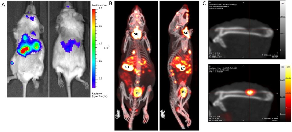
Fig.1 Metastatic prostate cancer model labeled with firefly luciferase (A) and NIS (B).1,4
FAQs about In Vivo Efficacy Evaluation
Q: Which tumor model should I choose for my ADC efficacy study?
A: Model selection depends on your development stage and scientific questions. For rapid initial screening with reproducible growth kinetics, we recommend cell-derived xenograft (CDX) models using our curated 70+ cell line panel. For translational validation with higher clinical predictivity, patient-derived xenograft (PDX) models preserve parental tumor characteristics more faithfully. If your ADC relies on Fc-mediated effector functions (ADCC, ADCP), syngeneic models in immunocompetent hosts are essential. For physiological tumor microenvironment contexts, GEMMs offer spontaneous tumorigenesis in native tissue compartments. We routinely help clients design tiered efficacy programs that progress through multiple model categories.
Q: How is tumor growth inhibition (TGI) calculated and interpreted in ADC efficacy studies?
A: TGI is expressed as a percentage using the formula: %TGI = (1 - ΔT_treated / ΔT_control) × 100, where ΔT represents the change in mean tumor volume over the observation period. A TGI greater than 50% is generally considered evidence of biologically meaningful activity, while TGI exceeding 80% typically indicates strong efficacy warranting advancement. We also report tumor regression events (partial or complete) and growth delay metrics (time for treated tumors to reach a predefined volume threshold relative to controls) to provide a comprehensive quantitative efficacy profile.
Q: What pharmacodynamic biomarkers do you measure to confirm ADC mechanism of action?
A: Our biomarker panel is tailored to your ADC's payload class and mechanism of action. For DNA-damaging payloads (e.g., PBD dimers, calicheamicins, topoisomerase I inhibitors), we quantify γ-H2AX (DNA double-strand breaks), phosphorylated ATM/ATR (damage response activation), and cleaved caspase-3 (apoptosis execution). For tubulin-acting payloads (e.g., auristatins, maytansinoids), we measure phospho-histone H3 (mitotic arrest) and mitotic spindle abnormalities. Additionally, we assess target antigen modulation, proliferation suppression (Ki-67 reduction), and immune cell infiltration (CD8+ T cells, NK cells) for Fc-effector characterization. All biomarkers are analyzed by quantitative IHC with digital pathology scoring, complemented by western blot or RT-qPCR as needed.
Q: Can you perform imaging-monitored efficacy studies to reduce animal numbers?
A: Yes. Our imaging suite includes an IVIS Spectrum system for bioluminescence imaging (BLI) of luciferase-expressing tumor models, enabling longitudinal tumor burden tracking in the same animals over the entire study duration. This approach substantially reduces inter-subject variability, decreases total animal usage in alignment with the 3Rs principles, and generates rich kinetic datasets suitable for PK/PD modeling integration. For orthotopic and GEMM applications where BLI is not applicable, we offer high-frequency ultrasound and small-animal MRI for serial anatomic tumor volumetry.
Q: How do you ensure that observed efficacy is specifically attributable to target antigen binding rather than non-specific accumulation?
A: We employ multiple orthogonal specificity controls depending on the model system. In CDX studies, we utilize CRISPR-engineered isogenic pairs — target antigen-knockout versus wild-type cells derived from the same parental line — grown in parallel to definitively attribute differential efficacy to antigen-specific binding versus passive EPR-mediated accumulation. Alternatively, we can include a non-binding isotype-control ADC at matched DAR and dosing to establish the baseline of non-targeted activity. In PDX studies, we correlate efficacy magnitude with quantitative target antigen expression level measured by IHC or flow cytometry across individual tumors, generating expression-response relationships that further support target-dependent activity attribution.
ADC Evaluation Solutions
Related Products
Related Resources

References:
1. Lyons, Scott K., Dennis Plenker, and Lloyd C. Trotman. "Advances in preclinical evaluation of experimental antibody-drug conjugates." Cancer Drug Resistance 4 (2021): 745-754. https://doi.org/10.20517/cdr.2021.37.
2. Liu, Yihan, Wantao Wu, Changjing Cai, Hao Zhang, Hong Shen, Ying Han, et al. "Patient-derived xenograft models in cancer therapy: technologies and applications." Signal Transduction and Targeted Therapy 8, no. 160 (2023). https://doi.org/10.1038/s41392-023-01419-2.
3. Long, Rou, Hanrong Zuo, Guiyang Tang, Chaohui Zhang, Xinru Yue, Jinsai Yang, Xinyu Luo, Yuqi Deng, Jieya Qiu, Jiale Li, and Jianhong Zuo. "Antibody-drug conjugates in cancer therapy: applications and future advances." Frontiers in Immunology 16 (2025): 1516419. https://doi.org/10.3389/fimmu.2025.1516419.
4. Distributed under Open Access License CC BY 4.0, without modification.
For Research Use Only. NOT FOR CLINICAL USE.
Online Inquiry
Welcome! For price inquiries, please feel free to contact us through the form on the left side. We will get back to you as soon as possible.
