- Home
- Resources
- Featured Solutions
- Universal Therapeutic Conjugate (UTC) Development Solution
- Antibody-Antibiotic Conjugate (AAC) Development Solution
- Bacterial Infection related Antibody Discovery
- S. aureus Antibody Development Services for AAC Research
- Anti-IsdB mAb Discovery: Preclinical S. aureus AAC Support
Anti-IsdB mAb Discovery: Preclinical S. aureus AAC Support
Iron-regulated surface determinant B (IsdB) is the primary hemoglobin receptor displayed on the Staphylococcus aureus cell surface — a conserved virulence factor that orchestrates heme-iron acquisition essential for bacterial survival and pathogenesis. Creative Biolabs provides a specialized anti-IsdB antibody discovery service designed to generate monoclonal antibody candidates that block heme-iron uptake, neutralize IsdB-mediated endothelial adhesion, and serve as high-precision targeting modules for antibody-antibiotic conjugate (AAC) development. Our multi-platform approach spans hybridoma, phage display, and individual-cell BCR repertoire profiling, supported by functional screening assays that prioritize growth inhibition and opsonophagocytic killing over binding affinity alone — delivering AAC-ready antibody candidates validated in iron-restricted in vitro conditions and in vivo infection models.
Inquire for Pre-clinical SupportOverview: IsdB as a High-Priority Antibacterial Target
IsdB is a ~72 kDa cell-wall-anchored protein encoded within the iron-regulated surface determinant (Isd) operon of S. aureus. Under the iron-restricted conditions characteristic of the mammalian host environment, IsdB is transcriptionally upregulated by the Fur (ferric uptake regulator) system and exposed on the bacterial surface, where it functions as the entry point of the Isd heme-transfer relay: IsdB captures hemoglobin and extracts heme via its tandem NEAT (NEAr Transporter) domains, then passes the heme cofactor sequentially through IsdA and IsdC to the membrane transporter IsdE/IsdF for cytoplasmic internalization. Once inside the bacterium, heme oxygenases IsdG and IsdI release elemental iron to sustain critical metabolic processes.
Three Therapeutic Angles for Anti-IsdB Antibodies
As a surface-exposed virulence factor with no functional homolog in humans, IsdB offers multiple intervention points for antibody-based strategies:
- • Heme-Iron Starvation: Antibodies binding the NEAT2 heme-binding pocket can directly block hemoglobin capture and heme extraction, depriving the pathogen of iron and inhibiting growth in the host environment.
- • Endothelial Adhesion Blockade: IsdB interacts with von Willebrand factor (vWF) to promote bacterial adhesion to activated endothelial cells — a critical step in infective endocarditis and metastatic infection. Anti-IsdB antibodies can disrupt this interaction.
- • AAC Internalization Receptor: IsdB undergoes active internalization upon antibody binding, making it an ideal receptor for delivering antibiotic payloads into intracellular S. aureus reservoirs via AAC technology.
IsdB vs. Other S. aureus Antigen Targets
| Feature | IsdB (Heme Receptor) | SpA (Protein A) | WTA (Wall Teichoic Acid) | ClfA (Clumping Factor A) |
|---|---|---|---|---|
| Biological Function | Hemoglobin binding & heme-iron acquisition | Fcγ & VH3 BCR binding (immune evasion) | Cell division & phage receptor | Fibrinogen binding & platelet aggregation |
| Surface Exposure | Upregulated in host (iron-limited) | Constitutive | Constitutive, abundant | Stationary phase predominant |
| Internalization Upon Ab Binding | Yes — enables intracellular AAC delivery | Limited | Unknown | Yes (fibrinogen-dependent) |
| Conservation Across Strains | High (MSSA & MRSA) | High | Serotype-dependent | Moderate |
| AAC Suitability | High (internalizing + pathogen-specific) | High (94 pM affinity demonstrated) | Moderate | Moderate |
Overcoming the Complexities of IsdB-Targeted Antibody Discovery
Despite its compelling target profile, IsdB presents several unique challenges that demand specialized discovery strategies beyond standard antibacterial antibody workflows:
- ▶ Dual-Receptor Redundancy: S. aureus expresses two surface hemoglobin receptors — IsdB and IsdH — that share approximately 65% sequence identity in their NEAT domains. IsdH can partially compensate for IsdB blockade, meaning antibodies targeting only IsdB may be insufficient for complete heme-acquisition inhibition in in vivo models.
- ▶ Non-Protective Immune Imprinting: Prior S. aureus exposure in human populations generates pre-existing IsdB-reactive antibodies predominantly targeting the non-protective NEAT1 domain rather than the heme-binding NEAT2 domain. Antibody discovery programs must actively exclude NEAT1-dominant clones to avoid selecting non-functional binders.
- ▶ Conformational Epitope Complexity: NEAT1 and NEAT2 perform distinct functions — NEAT1 mediates initial hemoglobin recognition while NEAT2 executes heme extraction. Antibodies targeting different domain interfaces produce divergent functional outcomes, requiring structural epitope characterization to distinguish growth-inhibitory binders from non-functional ones.
- ▶ Functional Assay Stringency: Conventional ELISA-based binding screens cannot distinguish antibodies that block heme acquisition from those that merely bind IsdB. Iron-limited growth conditions with hemoglobin as the sole iron source — coupled with opsonophagocytic killing (OPK) and vWF adhesion assays — are required for meaningful candidate triaging.
Our Targeted Anti-IsdB Antibody Discovery Solutions
Creative Biolabs has developed an IsdB-focused antibody discovery framework that integrates structural biology insights, dual-receptor screening, and functional prioritization to deliver AAC-ready candidates:
| Discovery Strategy | Technical Challenges Solved | Analytical Support & Validation |
|---|---|---|
|
Primary Strategy Dual-Target Heme-Blockade Immunization Immunogen design incorporating both IsdB NEAT2 and IsdH NEAT3 heme-binding pockets to generate cross-reactive antibodies that simultaneously block both hemoglobin receptors. |
• Eliminates IsdH-mediated compensation for IsdB blockade. • Targets structurally conserved heme-binding pocket residues (75% identity between IsdB NEAT2 and IsdH NEAT3). • Prevents bacterial iron acquisition even under high hemoglobin conditions. |
• Surface Plasmon Resonance (SPR): Competitive heme-binding inhibition kinetics for both IsdB and IsdH. • Isothermal Titration Calorimetry (ITC): Direct measurement of heme displacement thermodynamics. |
|
Epitope-Focused NEAT2-Domain Selective Screening Differential counter-screening against recombinant NEAT1 and NEAT2 domains to preferentially isolate antibodies targeting the protective heme-binding NEAT2 domain while excluding NEAT1-dominant clones. |
• Overcomes non-protective immune imprinting from prior S. aureus exposure. • Avoids selection of NEAT1-only binders that fail to block heme acquisition. • Enriches for clones with in vivo protective potential. |
• Differential ELISA/SPR: Parallel binding measurement against NEAT1 and NEAT2 recombinant proteins. • Hb-Dependent Growth Inhibition: MRSA strain panel tested in iron-limited medium with hemoglobin as sole iron source. |
|
Structural Epitope Binning by X-Ray Crystallography High-resolution co-crystal structures of antibody Fab:IsdB NEAT2 complexes to map epitope location relative to the heme-binding pocket, CDR3 engagement geometry, and domain interface coverage. |
• Distinguishes heme-pocket competitors from allosteric or distal binders. • Identifies CDR3 loop geometries that penetrate the heme-binding cavity for direct steric blockade. • Guides affinity maturation toward functionally relevant paratope residues. |
• X-Ray Crystallography: Fab:NEAT2 co-crystal structures at ≥2.0 Å resolution. • Alanine Scanning Mutagenesis: Functional epitope mapping to identify critical contact residues. |
|
Functional OPK & vWF Adhesion Dual Functional Assay Prioritizes antibody candidates through parallel opsonophagocytic killing assays and vWF-mediated endothelial adhesion blocking in primary human endothelial cell monolayers. |
• Ensures antibody candidates deliver both antibacterial effector function and anti-adhesion activity. • Identifies clones effective against diverse clinical MRSA/MSSA isolates. • Provides functional data directly translatable to in vivo efficacy prediction. |
• OPK Assay: Differentiated HL-60 phagocytes with complement, quantified by CFU enumeration. • Endothelial Adhesion Assay: Primary HUVEC monolayers activated with TNF-α, fluorescently labeled bacteria. |
Personalized Anti-IsdB Antibody Discovery Workflow
Our integrated workflow spans antigen design through AAC compatibility assessment, with functional data driving go/no-go decisions at each stage:
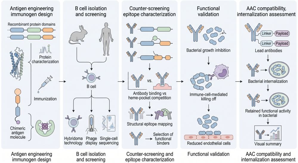
Phase 1: Antigen Engineering & Immunogen Design
We produce recombinant IsdB NEAT1, NEAT2, full-length IsdB, and IsdH NEAT3 proteins with confirmed heme-binding activity. For dual-target programs, chimeric immunogens incorporating conserved heme-pocket epitopes from both IsdB and IsdH are designed to elicit cross-reactive antibody responses. Each antigen batch undergoes biophysical characterization (SEC-MALS, CD spectroscopy) and functional validation by heme-titration SPR before immunization.
Phase 2: Multi-Platform B Cell Isolation & Screening
B cells are sourced from immunized animals (mouse, rat, rabbit, or camelid for VHH/single-domain antibodies) or from human donors with documented anti-S. aureus serology. Hybridoma fusion, phage display library construction, and individual-cell BCR repertoire profiling are deployed in parallel or as complementary approaches depending on program goals. Primary screening uses NEAT2-specific ELISA to pre-filter for heme-domain reactivity.
Phase 3: Dual-Target Counter-Screening & Epitope Characterization
Candidate antibodies are tested for cross-reactivity against IsdH NEAT3 and counter-screened against NEAT1 to exclude non-functional domain binders. Lead clones are subjected to SPR-based heme competition assays and, for top candidates, Fab:NEAT2 co-crystallography to map epitope location relative to the heme-binding cavity. Alanine scanning mutagenesis of critical NEAT2 contact residues further refines the functional epitope map.
Phase 4: Functional Validation in Physiologically Relevant Assays
Lead antibodies are ranked by functional performance, not binding affinity alone. Key assays include: (a) hemoglobin-dependent growth inhibition of MRSA and MSSA clinical isolates in iron-limited medium, (b) opsonophagocytic killing using differentiated HL-60 effector cells with human complement, and (c) vWF-mediated endothelial adhesion blockade on TNF-α-activated primary HUVEC monolayers. Antibodies demonstrating activity in at least two of three assays advance to the AAC compatibility stage.
Phase 5: AAC Conjugation Compatibility & Internalization Assessment
Functional lead antibodies are evaluated for AAC conjugation feasibility: assessment of available lysine/cysteine residues for linker chemistry, confirmation of antibody-mediated IsdB internalization in MRSA using pH-sensitive fluorophore conjugates, and validation that conjugated antibodies retain heme-blockade and OPK activity. A final report summarizes antibody sequences, epitope maps, functional data, conjugation compatibility, and recommendations for linker-payload pairing for pre-clinical AAC evaluation.
Technology Platforms for Anti-IsdB Antibody Discovery
Our discovery platforms are purpose-built for IsdB biology, integrating structural knowledge of the NEAT domain system with functional screening rigor:
1. Recombinant IsdB/IsdH Multi-Domain Antigen Platform
A dedicated recombinant protein production suite for IsdB NEAT1, IsdB NEAT2, full-length IsdB, and IsdH NEAT3. All proteins are produced in E. coli or mammalian expression systems with confirmed heme-binding activity validated by UV-visible spectroscopy and SPR heme titration. This platform enables differential screening strategies that distinguish NEAT2-specific from NEAT1-dominant antibody responses.
- • Heme-Loaded & Apo Forms: Antigens produced in both heme-bound and heme-free states for epitope accessibility screening.
- • Chimeric Immunogens: IsdB-IsdH fusion constructs incorporating conserved heme-pocket residues for dual-target antibody generation.
- • Biotinylated Probes: Site-specific biotinylation for SPR and Octet immobilization without masking the heme-binding interface.
2. Multi-Format B Cell Antibody Discovery Suite
A comprehensive antibody discovery arsenal spanning classical hybridoma technology, immune and synthetic phage display libraries, and high-throughput individual-cell BCR repertoire profiling. This multi-format flexibility allows program-specific selection of the optimal discovery modality based on target biology, desired antibody format, and timeline requirements.
- • Hybridoma: Classical mouse/rat immunization with IsdB NEAT2 antigen, PEG-mediated fusion, HAT selection, and clonal expansion.
- • Phage Display: Immune library from immunized animals or synthetic library panning against recombinant IsdB NEAT2 with heme-competition elution.
- • Individual-cell BCR Repertoire Profiling: Direct paired VH/VL recovery from antigen-specific B cells sorted by NEAT2-fluorophore probes.
3. Heme-Blockade Functional Screening Platform
A specialized functional screening cascade designed to distinguish antibodies that actively block heme acquisition from those that merely bind IsdB. The platform operates under physiologically relevant iron-limited conditions with hemoglobin as the sole iron source, directly measuring bacterial growth inhibition rather than binding signal.
- • Hb-Dependent Growth Assay: MRSA/MSSA panel cultured in iron-depleted RPMI + 10 μM hemoglobin, OD600 monitoring over 24 hours.
- • SPR Heme Competition: Quantitative measurement of antibody-mediated blockade of heme binding to immobilized NEAT2.
- • OPK Functional Assay: Differentiated HL-60 effector cells + human complement, CFU quantification at 2 hours.
4. AAC Conjugation & Internalization Assessment Platform
An integrated bioconjugation and cell-based evaluation suite that determines whether functional anti-IsdB antibodies are suitable for AAC development. This platform assesses conjugation chemistry compatibility, confirms antibody-dependent internalization through IsdB, and validates that the conjugated antibody retains target binding and functional activity for pre-clinical AAC evaluation.
- • Conjugation Feasibility: Assessment of solvent-accessible lysine/cysteine residues, interchain disulfide integrity, and aggregation propensity post-conjugation.
- • Internalization Assay: pHrodo-labeled antibody tracking in live MRSA, confocal microscopy confirmation of phagolysosomal delivery.
- • Post-Conjugation Functional Retention: Re-testing of heme blockade and OPK activity after linker-payload conjugation.
Why Choose Our Anti-IsdB Antibody Discovery Services?
IsdB Biology-Driven Discovery Design
Our platform is built on deep structural and functional knowledge of the Isd heme-transfer system — from NEAT domain architecture to heme-extraction kinetics — ensuring every screening decision is informed by the underlying biology rather than generic antibody discovery workflows.
Dual-Receptor Coverage Against IsdB and IsdH
We address the IsdH compensation problem head-on with immunogens and screening strategies designed to identify antibodies that cross-neutralize both hemoglobin receptors, maximizing growth-inhibitory potency against diverse S. aureus clinical isolates.
AAC-Optimized Antibody Delivery
Antibody candidates are evaluated for AAC compatibility from the outset — internalization efficiency, conjugation chemistry accessibility, and post-conjugation functional retention are assessed before candidate nomination, not as an afterthought.
Function-First Candidate Prioritization
Growth inhibition in iron-restricted conditions, opsonophagocytic killing activity, and endothelial adhesion blockade — not binding affinity alone — drive our candidate ranking. This functional prioritization ensures that antibodies entering AAC development have demonstrated antibacterial mechanisms of action.
Research Insights: IsdB as an Antibacterial Antibody Target
A growing body of structural and functional evidence positions IsdB as a high-priority target for antibacterial antibody development. According to Alfeo et al. (2021), IsdB functions not only as a heme-iron acquisition receptor but also as a von Willebrand factor (vWF)-binding adhesin that mediates S. aureus attachment to activated endothelial cells — a critical early step in infective endocarditis. Patient-derived IsdB-reactive IgG from endocarditis cases nearly completely blocked bacterial adhesion, demonstrating that antibodies targeting IsdB can exert dual antibacterial mechanisms: nutrient starvation and adhesion blockade.
Structural & Functional Advances Supporting IsdB Targeting:
- • Dual-Receptor Polypeptide Fragment: Valenciano-Bellido et al. (2023) reported the first camelid-derived polypeptide fragment capable of simultaneously recognizing the heme-binding pockets of both IsdB NEAT2 and IsdH NEAT3 with nanomolar affinity. Crystal structures at 1.65–1.70 Å resolution revealed CDR2/CDR3 engagement geometry that directly competes with heme, and polypeptide fragment 6 inhibited growth of three MRSA clinical isolates (USA300, MRSA49, MRSA54) in hemoglobin-dependent iron-limited conditions — providing proof-of-concept for dual-receptor blockade.
- • NEAT Domain Division of Labor: A 2025 study used time-resolved X-ray solution scattering (TR-XSS) to capture the real-time structural dynamics of IsdB interacting with hemoglobin. Their work established that NEAT1 mediates initial hemoglobin recognition while NEAT2 executes heme extraction, and revealed that the α-subunit binding step represents a previously unappreciated intervention point for antibody blockade — expanding the accessible epitope space beyond the heme-binding cavity itself.
- • AAC Design Framework: Cavaco et al. (2022) comprehensively reviewed AAC development principles including antibody selection criteria, linker chemistry (protease-cleavable Val-Cit vs. non-cleavable), payload conjugation strategies, and pharmacokinetic considerations — providing a design blueprint for translating anti-IsdB antibodies into functional AAC candidates.
These complementary insights from structural biology, antibody engineering, and conjugate design collectively validate IsdB as a premier target for AAC-enabled antibacterial strategies in the pre-clinical space.
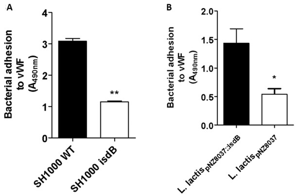
Fig.1 Adhesion of S. aureus and IsdB-expressing L. lactis to immobilized vWF.1, 4
FAQs about Anti-IsdB Antibody Discovery
Q: What makes IsdB a compelling target compared to other S. aureus surface antigens?
A: IsdB offers three distinct therapeutic angles in a single target: (1) blocking heme-iron acquisition starves the pathogen of an essential nutrient, (2) disrupting vWF-mediated endothelial adhesion prevents tissue colonization, and (3) antibody-induced IsdB internalization enables AAC payload delivery to intracellular bacterial reservoirs. Unlike Protein A, which primarily serves immune evasion, IsdB targeting directly compromises bacterial metabolism and survival.
Q: How does your approach address the IsdH redundancy problem in heme acquisition?
A: Our dual-target strategy incorporates immunogen designs that present conserved heme-binding pocket epitopes shared between IsdB NEAT2 and IsdH NEAT3 (approximately 75% identity within the heme-binding cavity). Antibody candidates are screened for cross-reactivity against both receptors, and only clones demonstrating competitive inhibition of heme binding to both IsdB and IsdH are advanced. This ensures that the final antibody cannot be bypassed by IsdH upregulation.
Q: What antibody formats are available for anti-IsdB discovery programs?
A: We support multiple discovery formats including conventional IgG (mouse, rat, rabbit via hybridoma), Camelid-derived heavy-chain variable domain antibodies (via phage display), and fully human antibodies (via individual-cell BCR repertoire profiling from convalescent donors or phage display). The choice of format depends on your program goals: polypeptide fragments offer superior tissue penetration and heme-pocket accessibility, while full IgG formats provide Fc-mediated effector functions such as opsonophagocytic killing.
Q: Are the anti-IsdB antibodies you discover directly compatible with AAC conjugation?
A: Yes. Antibody candidates are evaluated for AAC compatibility during Phase 5 of our workflow, including assessment of accessible conjugation sites, post-conjugation functional retention (heme blockade + OPK), and internalization efficiency. We provide recommendations for linker chemistry (e.g., protease-cleavable Val-Cit linkers for lysosomal release) and payload pairing based on the antibody's internalization kinetics and the target bacterial species' antibiotic sensitivity profile.
Q: What S. aureus strains are included in your functional validation panel?
A: Our standard validation panel includes at least three clinically relevant MRSA strains (e.g., USA300, MRSA49/54, or client-specified临床 isolates) and at least one MSSA reference strain. Growth inhibition is tested in iron-limited RPMI medium supplemented with 10 μM human hemoglobin as the sole iron source — conditions that mimic the host environment and require functional IsdB-mediated heme acquisition for bacterial survival. Additional custom strain panels can be accommodated upon request.
Related S. aureus related Antibody Discovery
Related Resources

References:
1. Alfeo, Mariangela J., et al. "Staphylococcus aureus iron-regulated surface determinant B (IsdB) protein interacts with von Willebrand factor and promotes adherence to endothelial cells." Scientific Reports 11 (2021): 22799. https://doi.org/10.1038/s41598-021-02065-w
2. Valenciano-Bellido, Sandra, et al. "Targeting hemoglobin receptors IsdH and IsdB of Staphylococcus aureus with a single VHH antibody inhibits bacterial growth." Journal of Biological Chemistry 299.9 (2023): 104927. https://doi.org/10.1016/j.jbc.2023.104927
3. Cavaco, Marco, Miguel A. R. B. Castanho, and Vera Neves. "The Use of Antibody-Antibiotic Conjugates to Fight Bacterial Infections." Frontiers in Microbiology 13 (2022): 835677. https://doi.org/10.3389/fmicb.2022.835677
4. All references distributed under Open Access License CC BY 4.0, without modification.
For Research Use Only. NOT FOR CLINICAL USE.
Online Inquiry
Welcome! For price inquiries, please feel free to contact us through the form on the left side. We will get back to you as soon as possible.
