- Home
- Resources
- Featured Solutions
- Universal Therapeutic Conjugate (UTC) Development Solution
- Antibody-Antibiotic Conjugate (AAC) Development Solution
- Bacterial Infection related Antibody Discovery
- S. aureus Antibody Development Services for AAC Research
- Anti-PBP-2a mAb Discovery: Preclinical MRSA AAC Support
Anti-PBP-2a mAb Discovery: Preclinical MRSA AAC Support
Penicillin-binding protein 2a (PBP-2a), the 78 kDa transpeptidase encoded by the mecA gene, is the defining molecular driver of broad-spectrum β-lactam resistance across all MRSA strains. Unlike the four native staphylococcal PBPs, PBP-2a maintains catalytic activity at antibiotic concentrations that fully saturate PBP1–4, making it an exceptional antibody target for next-generation anti-MRSA strategies. Creative Biolabs provides comprehensive antibody-antibiotic conjugate (AAC)-compatible anti-PBP-2a antibody discovery services. Our multi-platform approach combines hybridoma, phage display, and single B cell cloning technologies with rigorous opsonophagocytic killing (OPK) validation, delivering well-characterized antibody candidates ready for covalent conjugation to potent anti-staphylococcal payloads during preclinical evaluation.
Inquire for Preclinical SupportOverview: The PBP-2a Resistance Paradigm as an Antibody Target
Methicillin-resistant Staphylococcus aureus (MRSA) remains one of the most clinically consequential multidrug-resistant pathogens worldwide. Its resistance phenotype is conferred by the acquisition of the mecA gene within the staphylococcal cassette chromosome mec (SCCmec), which encodes PBP-2a—a unique penicillin-binding protein absent from methicillin-susceptible strains. PBP-2a catalyzes the transpeptidation step of peptidoglycan cross-linking with a remarkably low affinity for virtually all β-lactam antibiotics, effectively bypassing the blockade imposed on the four native PBPs and enabling cell wall biosynthesis to proceed uninterrupted at therapeutic antibiotic concentrations.
Structural Architecture of PBP-2a
Structurally, the 78 kDa PBP-2a comprises four distinct domains organized around its membrane-proximal anchor and extracellular catalytic machinery:
| Domain | Approximate Residues | Functional Role |
|---|---|---|
| Transmembrane Anchor | 1–23 | Hydrophobic N-terminal α-helix that tethers PBP-2a to the cytoplasmic membrane, positioning the extracellular catalytic domains at the peptidoglycan synthesis interface. |
| N-terminal Extension (NTE) | 27–326 | Non-catalytic domain that serves as a regulatory scaffold. The allosteric site within the NTE receives the stem peptide signal from nascent peptidoglycan, triggering a conformational relay that opens the active site cleft for substrate access. |
| Non-penicillin-binding (nPB) Domain | 327–642 | Structurally conserved domain connecting the allosteric sensor to the transpeptidase domain. Forms a large cleft with the TP domain that contributes to substrate selectivity and antibiotic discrimination. |
| C-terminal Transpeptidase (TP) Domain | 643–668 | Contains the catalytic serine-active site (S403-T404-Q405-K406 motif) responsible for DD-transpeptidation. The closed, occluded active site conformation is the structural basis for β-lactam evasion—antibiotics cannot efficiently access the catalytic serine unless the allosteric gate is opened. |
This intricate allosteric regulation—whereby the NTE senses cell wall maturation and induces a conformational transition that opens the TP domain for catalysis—defines PBP-2a as both the critical determinant of MRSA viability under β-lactam pressure and a structurally validated antibody target on the extracellular surface.
Challenges in Targeting the PBP-2a Allosteric Enzyme
Although PBP-2a is exposed on the outer face of the cytoplasmic membrane and is universally conserved across MRSA isolates, several intrinsic features complicate antibody development:
- ▶ Conformationally Gated Active Site: The TP domain active site is predominantly in a closed, ligand-inaccessible conformation. Antibodies must either trap the open state or target the allosteric NTE to block the conformational switch that enables catalysis.
- ▶ Low Immunogenic Presentation on Intact Bacteria: As a membrane-associated protein with a compact extracellular domain, PBP-2a presents fewer solvent-exposed epitopes compared to abundant cell wall-anchored surface proteins, potentially yielding lower-affinity polyclonal responses from standard immunization protocols.
- ▶ Limited Therapeutic Window of Monotherapy: Even potent anti-PBP-2a monoclonal antibodies achieve approximately 1–2 log10 bacterial burden reduction in murine sepsis models—clinically meaningful but rarely sterilizing as a standalone intervention. This motivates the development of AAC-formatted candidates in which the antibody delivers a bactericidal antibiotic payload directly to the MRSA cell surface.
- ▶ Conjugation-Site Accessibility Assessment: Mapping antibody surface lysine residues that remain accessible after PBP-2a binding requires iterative HDX-MS and computational solvent accessibility analysis to ensure AAC conjugation does not compromise target engagement in in vivo infection models.
Multi-Platform Anti-PBP-2a Antibody Discovery Solutions
Our integrated antibody discovery pipeline addresses each challenge through platform-matched strategies, from recombinant antigen design to AAC-compatible candidate selection:
| Discovery Platform | Technical Challenges Resolved | Key Validation Endpoints |
|---|---|---|
|
Core Platform Recombinant Domain Immunization — Hybridoma Immunization with recombinant PBP-2a ectodomain or the isolated allosteric NTE fragment, screened against full-length PBP-2a expressed on S. aureus protoplasts, to capture conformation-sensitive binders. |
• Overcomes low immunogenicity by delivering high-concentration recombinant antigen enriched for the extracellular domain. • Targets the NTE allosteric site to block the conformational switch without requiring active-site access. • Protoplast-based screening ensures antibodies bind the native membrane-associated conformation. |
• Whole-cell ELISA against isogenic MRSA and MSSA pairs to confirm PBP-2a specificity. • Latex agglutination for rapid PBP-2a binding confirmation. • Competition ELISA with β-lactam probes to assess active-site proximity. |
|
Complementary Phage Display Panning on Conformationally Stabilized PBP-2a Naive and immune phage display libraries panned against PBP-2a captured in the allosterically primed open conformation using a covalent cephalosporin intermediate. |
• Captures antibodies specific to the open, catalytically active TP conformation. • Enables selection of human or humanized frameworks directly, bypassing murine immunogenicity. • Rapid affinity maturation through CDR-targeted mutagenesis libraries. |
• Biolayer interferometry (BLI) for real-time binding kinetics (kon, koff, KD). • HDX-MS epitope mapping to locate the antibody footprint on the PBP-2a surface. • Allosteric modulation assay: monitoring TP activity in the presence and absence of antibody. |
|
High-Throughput Single B Cell Cloning & Functional Screening Fluorescence-labeled PBP-2a ectodomain tetramers to isolate antigen-specific memory B cells from immunized animals, followed by individual-cell RT-PCR and recombinant IgG expression. |
• Preserves native heavy-light chain pairing for optimal affinity and expression. • Parallel functional screening eliminates non-neutralizing binders at the clonal level. • Generates sequence-defined recombinant mAbs suitable for subsequent humanization. |
• Flow cytometry sorting with PBP-2a tetramer and S. aureus whole-cell counter-screening. • Miniaturized OPK assay with 96-well format fresh human neutrophil readout. • Sequencing + recombinant expression for each positive clone. |
|
AAC Integration OPK Functional Validation & AAC Compatibility Assessment Quantitative opsonophagocytic killing assays in whole human blood, combined with post-conjugation binding retention ELISA, to select lead candidates that maintain target engagement after antibiotic payload attachment. |
• Directly measures the functional consequence of antibody binding—neutrophil-mediated bacterial clearance. • Screens for AAC compatibility before committing to conjugate synthesis. • Identifies the Fc isotype that maximizes OPK activity (IgG1 vs. IgG3) to inform Fc engineering. |
• Whole-blood OPK assay: % killing at 2 h with fresh human neutrophils and baby rabbit complement. • Post-conjugation ELISA: antigen-binding retention after model linker-payload attachment. • Antibiotic synergy checkerboard: mAb + vancomycin or daptomycin fractional inhibitory concentration (FIC) index. |
Integrated Anti-PBP-2a Antibody Discovery Workflow
Our standardized five-phase process delivers AAC-ready anti-PBP-2a antibody candidates backed by structural, functional, and conjugation-compatibility data:
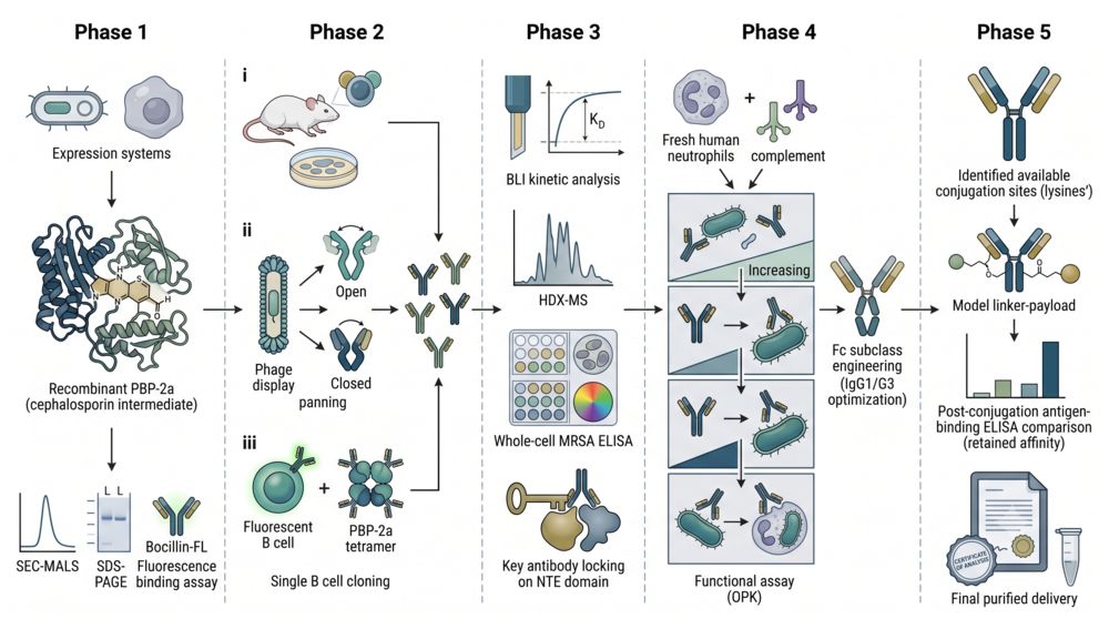
Phase 1: Antigen Design & Conformational Stabilization
We express recombinant PBP-2a ectodomain (residues 27–668) and the isolated NTE (residues 27–326) in E. coli or mammalian systems. For conformation-specific campaigns, PBP-2a is trapped in the open state using a covalent cephalosporin intermediate. Quality control includes SEC-MALS for monodispersity, SDS-PAGE for purity, and a cephalosporin bocillin-FL binding assay to confirm catalytic competence.
Phase 2: Multi-Platform Antibody Generation
Three parallel tracks run simultaneously: (i) murine hybridoma from animals immunized with recombinant PBP-2a ectodomain or NTE; (ii) phage display libraries panned against allosterically primed and resting PBP-2a; (iii) single B cell cloning using fluorescent PBP-2a tetramers. This redundancy ensures at least one platform produces high-affinity, conformationally selective binders targeting diverse epitopes across the NTE, nPB, and TP domains.
Phase 3: Binding Kinetics & Epitope Characterization
Lead candidates undergo BLI-based kinetic analysis (KD < 10 nM threshold), HDX-MS epitope binning, and whole-cell MRSA ELISA to confirm native binding. Antibodies recognizing the NTE allosteric domain are prioritized for their potential to lock PBP-2a in the closed, inactive conformation. Competitive binding against a β-lactam-biotin probe quantifies active-site proximity.
Phase 4: Functional Validation by Opsonophagocytic Killing
The functional cornerstone of candidate selection: quantitative whole-blood OPK assays using fresh human neutrophils and baby rabbit complement. Each candidate is tested at three concentrations (1–100 μg/mL) against at least two genetically distinct MRSA strains. Candidates achieving ≥50% killing at ≤10 μg/mL advance to AAC compatibility testing. Fc engineering (IgG1/G3 subclass optimization) is applied to maximize FcγR-mediated uptake.
Phase 5: AAC-Ready Candidate Delivery
Top-ranked candidates undergo computational conjugation-site analysis to identify lysine or cysteine residues that remain solvent-accessible after PBP-2a binding. A model linker-payload is conjugated at lysine residues, and post-conjugation antigen-binding ELISA confirms retained affinity (>80% of unconjugated antibody). Final delivery includes purified antibody, full characterization report, conjugation compatibility data, and sequence information for subsequent humanization.
Core Technology Platforms for PBP-2a Antibody Development
Each platform is configured to address the unique structural and functional demands of targeting a membrane-associated allosteric enzyme:
1. Recombinant PBP-2a Antigen Engineering Platform
We produce high-purity recombinant PBP-2a ectodomain with or without covalent allosteric priming. The antigen is validated by SEC-MALS for monodispersity and by bocillin-FL competition assay to confirm that the open conformation retains catalytic competence. The NTE-only construct is used in parallel immunization arms to enrich for allosteric-site-directed antibodies.
- • Conformational QC: Bocillin-FL binding and competition to verify active-site integrity.
- • NTE isolation: Domain-specific immunization to drive allosteric-site antibody enrichment.
- • Protoplast screening: Ensures isolated mAbs recognize native membrane-associated PBP-2a on live bacteria.
2. High-Throughput OPK Screening Platform
A standardized 96-well opsonophagocytic killing assay using fresh human polymorphonuclear neutrophils (PMNs) from healthy donors and baby rabbit complement. Each plate includes a no-antibody negative control, an irrelevant IgG isotype control, and a pooled human IVIG positive control. Bacterial survival is quantified by serial dilution plating at 0 and 2 hours, with killing expressed as log10 reduction.
- • Multi-strain testing: Tested against at least two genetically distinct MRSA lineages (e.g., ST239 and ST8/USA300).
- • Dose-response: Three concentrations per candidate to identify the OPK EC50.
- • Fc isotype comparison: IgG1 vs. IgG3 Fc formats evaluated side by side for OPK potency.
3. AAC Conjugation Compatibility Suite
Before investing in full conjugate synthesis, lead antibodies undergo computational solvent-accessible surface area (SASA) analysis to identify lysine residues that remain exposed after antigen docking. A model linker (non-cleavable SMCC or protease-cleavable val-cit-PAB) is conjugated at surface lysines, and the conjugate is tested by ELISA to confirm >80% antigen-binding retention. Hydrophobic interaction chromatography (HIC) confirms the absence of aggregation-prone high-DAR species.
- • Computational conjugation site mapping: SASA-based lysine ranking pre- and post-docking.
- • Model conjugate ELISA: Post-conjugation binding retention as a gate for full synthesis.
- • HIC aggregation screen: Rules out conjugation-induced aggregation at the candidate stage.
4. Antibody Humanization & Fc Engineering Platform
Murine lead candidates undergo CDR grafting onto human germline frameworks (IGHV1-69 or IGHV3-23 for heavy chains; IGKV1-39 or IGKV3-20 for light chains) with back-mutation guided by structural modeling. Fc engineering options include: (i) S239D/I332E mutations to enhance FcγRIIIa binding and OPK activity; (ii) M252Y/S254T/T256E (YTE) mutations to extend serum half-life; and (iii) L234A/L235A (LALA) mutations to ablate FcγR binding if complement-dependent OPK is the dominant clearance mechanism.
- • CDR grafting + back-mutation: Preserves affinity while minimizing immunogenicity risk.
- • OPK-optimized Fc: S239D/I332E for enhanced neutrophil-mediated clearance.
- • Half-life extension: YTE mutations for extended in vivo pharmacokinetic exposure in preclinical infection models.
Why Our Anti-PBP-2a Antibody Discovery Program
Structure-Guided Antigen Design
Our antigen strategy leverages the known allosteric mechanism of PBP-2a: by presenting the NTE domain in isolation or trapping the TP domain in the open conformation, we direct the immune repertoire toward functionally relevant epitopes that a conventional whole-protein immunization would miss. This structural awareness distinguishes our program from generic anti-bacterial antibody campaigns.
Functional Screening, Not Just Binding
Every candidate is tested in quantitative whole-blood OPK assays before entering the lead selection pipeline. Binding affinity (KD) is necessary but insufficient; only antibodies that drive neutrophil-mediated bacterial killing advance. This functional-first philosophy ensures the delivered candidates possess the biological activity required for AAC development.
AAC-Ready from Day One
Unlike standalone antibacterial antibody programs, our workflow integrates AAC compatibility assessment at every stage—from computational conjugation-site mapping before immunization to post-conjugation binding retention ELISA on lead candidates. The antibodies you receive are not merely anti-PBP-2a binders; they are pre-validated for covalent antibiotic delivery.
Comprehensive Preclinical Characterization
Every delivered candidate includes: HDX-MS epitope map, BLI kinetics (kon, koff, KD), whole-cell MRSA/MSSA specificity ELISA, whole-blood OPK dose-response (EC50), post-conjugation binding retention ratio, and antibiotic synergy FIC index. This data package supports immediate progression to in vivo MRSA infection model efficacy studies.
Research Insights: Anti-PBP-2a Antibodies in MRSA Immunotherapy
According to Saraiva et al. (2019), a murine monoclonal antibody targeting a conserved 88-amino-acid region of PBP-2a that encompasses the catalytic serine-active-site motif (STQK) provided significant protection in both prophylactic and therapeutic MRSA infection models. Their study established that anti-PBP-2a antibodies can function through a direct neutralization mechanism independent of host immune effector functions.
Key Findings from PBP-2a Antibody Research:
- • Direct Enzyme Neutralization: F(ab′)2 fragments lacking the Fc region retained protective efficacy, demonstrating that the antibody interferes with PBP-2a enzymatic function directly. This mechanism is particularly relevant for AAC design, as the antibody scaffold remains functional even when conjugation sterically constrains FcγR engagement.
- • Comparable Efficacy to Vancomycin: In a therapeutic treatment model (antibody administered 6 h post-infection), anti-PBP-2a mAb reduced kidney bacterial load by 96.2%, statistically equivalent to vancomycin (93.4%). The mAb-plus-vancomycin combination achieved 98.9% reduction, suggesting additive benefit from orthogonal mechanisms.
- • Broad Strain Coverage: Protection was demonstrated against two genetically distinct MRSA clones (BEC/ST239 and Iberian/ST247), indicating that the conserved active-site region allows cross-strain efficacy—a critical attribute for a universal anti-MRSA AAC agent.
These data position PBP-2a as a validated extracellular antibody target for MRSA and support the rationale for AAC development: an antibody that both neutralizes PBP-2a and delivers a bactericidal antibiotic payload to the bacterial surface may achieve the sterilizing activity that monotherapy alone cannot provide.
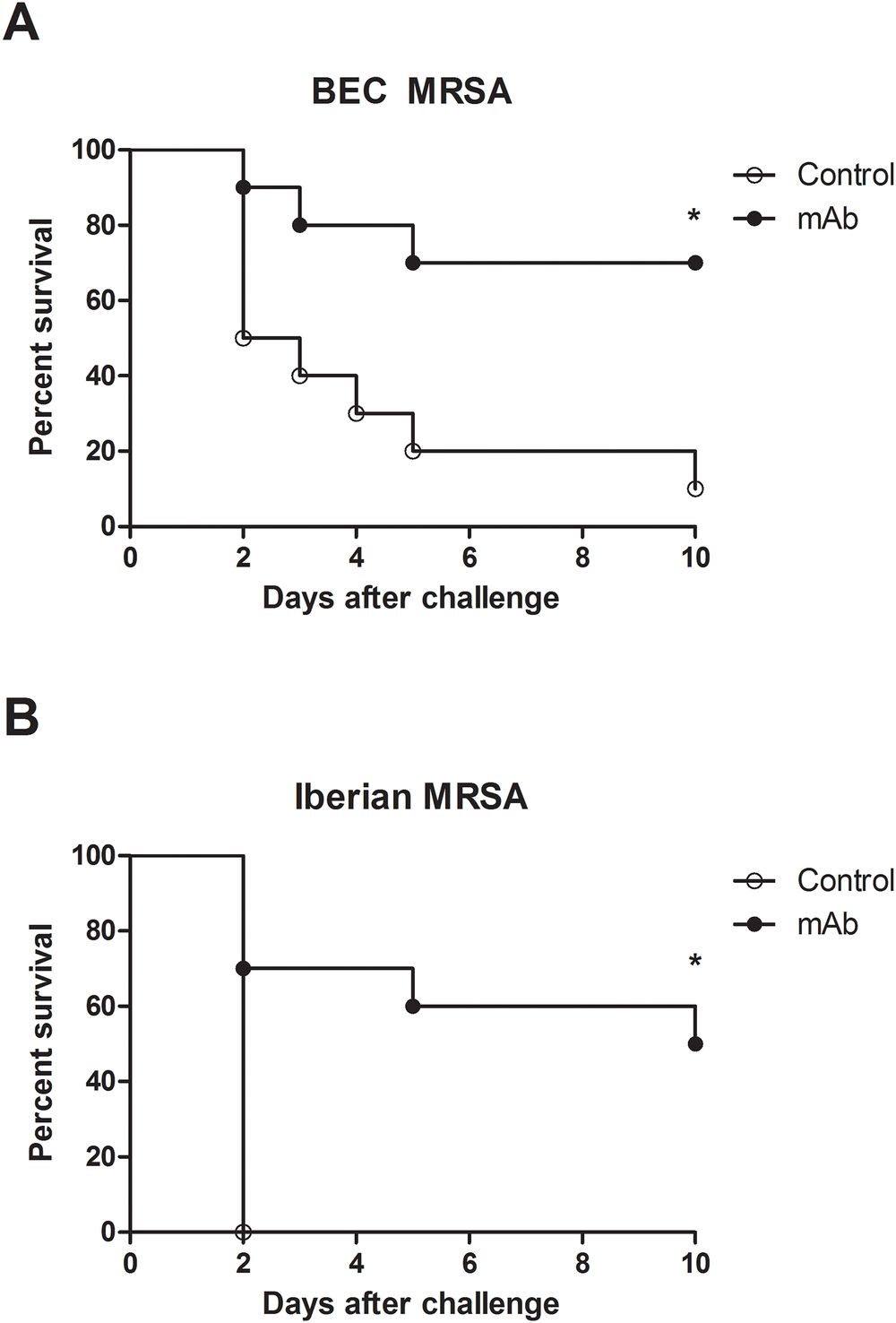
Fig.1 Prophylactic anti-PBP2a mAb efficacy against lethal BEC and Iberian MRSA challenge.1,5
FAQs about Anti-PBP-2a Antibody Discovery
Q: Why target PBP-2a rather than abundant cell wall-anchored surface proteins like Protein A or ClfA?
A: PBP-2a is the molecular determinant of broad-spectrum β-lactam resistance—it is absent from methicillin-susceptible S. aureus (MSSA) and universally present in all MRSA strains. An anti-PBP-2a AAC therefore selectively targets the resistant population while sparing the normal flora. Furthermore, antibodies binding the NTE allosteric domain can directly block the enzymatic activity that sustains cell wall synthesis under antibiotic pressure, adding a functional neutralization mechanism beyond simple opsonization.
Q: What is the advantage of combining anti-PBP-2a antibodies with AAC technology?
A: Anti-PBP-2a monotherapy achieves approximately 1–2 log10 bacterial reduction in preclinical models—significant but rarely sterilizing. Conjugating a bactericidal antibiotic payload (e.g., a rifamycin analog or glycopeptide derivative) to the antibody creates a dual-mechanism agent: the antibody neutralizes PBP-2a transpeptidase activity and delivers a high-local-concentration antibiotic payload to the MRSA surface, overcoming the pharmacokinetic limitations of systemic antibiotic administration at the infection site.
Q: Do you test antibody candidates against multiple MRSA strain backgrounds?
A: Yes. Our standard OPK validation panel includes at least two genetically distinct MRSA lineages (e.g., a hospital-associated ST239 clone and a community-associated USA300/ST8 clone). Whole-cell ELISA against isogenic MRSA/MSSA pairs confirms PBP-2a specificity. Additional strain testing can be expanded per project requirements to include regional clinical isolates of interest.
Q: How do you ensure the antibody remains functional after antibiotic conjugation?
A: Before full-scale AAC synthesis, our computational pipeline ranks all surface-exposed lysine residues on the antibody by SASA (solvent-accessible surface area) and proximity to the HDX-MS-defined PBP-2a epitope footprint. A model linker-payload is then conjugated, and post-conjugation binding is quantified by ELISA. Only antibodies retaining >80% of unconjugated binding affinity advance to full conjugate production, ensuring that your final AAC candidate maintains target engagement.
Q: What deliverables are included in a typical anti-PBP-2a antibody discovery project?
A: Each project delivers purified lead antibody (typically 3–5 candidates), a comprehensive characterization report (BLI kinetics, HDX-MS epitope map, whole-cell ELISA specificity, whole-blood OPK dose-response with EC50), model-conjugate binding retention data, antibiotic synergy FIC indices, and antibody sequences for downstream humanization. Additional in vivo efficacy testing in murine MRSA infection models is available as a follow-on service.
Related S. aureus related Antibody Discovery
Related Products
Related Resources

References:
1. Saraiva, Felipe Betoni, et al. "Monoclonal antibody anti-PBP2a protects mice against MRSA (methicillin-resistant Staphylococcus aureus) infections." PLoS ONE 14.11 (2019): e0225752. https://doi.org/10.1371/journal.pone.0225752
2. Cavaco, Marco, et al. "The Use of Antibody-Antibiotic Conjugates to Fight Bacterial Infections." Frontiers in Microbiology 13 (2022): 835677. https://doi.org/10.3389/fmicb.2022.835677
3. Speziale, Pietro, and Giampiero Pietrocola. "Monoclonal Antibodies Targeting Surface-Exposed and Secreted Proteins from Staphylococci." Vaccines 9.5 (2021): 459. https://doi.org/10.3390/vaccines9050459
4. Arêde, Pedro, et al. "The anti-repressor MecR2 promotes the proteolysis of the mecA repressor and enables optimal expression of β-lactam resistance in MRSA." PLoS Pathogens 8.7 (2012): e1002816. https://doi.org/10.1371/journal.ppat.1002816
5. Distributed under Open Access License CC BY 4.0, without modification.
For Research Use Only. NOT FOR CLINICAL USE.
Online Inquiry
Welcome! For price inquiries, please feel free to contact us through the form on the left side. We will get back to you as soon as possible.
