

Support
Creative Biolabs aims to help you achieve reproducible results by providing validated antibodies and technical support to facilitate your immunohistochemistry (IHC) projects. The technical background we can provide includes:
What is IHC?
IHC is the process of using antibodies to detect cellular proteins in tissue sections and covers anatomical, immunological, and biochemical techniques. IHC is a well-established, highly sensitive method for localizing antigens within cells or tissues with high resolution. Based on the in situ specific binding of appropriately labeled antibodies to their target antigens to image discrete components in tissue, IHC technology combines molecular detection with morphological characterization and can be used on formalin-fixed paraffin-embedded sections, frozen sections, and other cytological preparations. IHC is also used to clarify the differential diagnosis, overcoming the limitations of traditional hematoxylin and eosin analysis. In recent years, advances in antigen exposure techniques and sensitive visualization methods in preserved tissues have made IHC an indispensable tool in modern molecular diagnostic laboratories.
Principle of IHC
The basic principle of IHC is that the binding of antibodies to antigens is highly specific. First, specific chemical substances in tissues or cells are extracted as antigens, and then specific antibodies are obtained by immunizing animals, and antibodies can be used for the detection of similar antigenic substances in tissues or cells. Since antigen-antibody complexes are not easily observed, specific labels, such as fluorescein, enzymes, and isotopes, need to be used to visualize the binding sites of antigens and antibodies to detect unknown antigens. In situ hybridization (ISH), a variant of IHC, is similarly used to detect nuclear markers using probes complementary to genomic sequences.
Applications of IHC
- IHC in tumors
IHC is the process of using antibodies to detect proteins in cells within tissue sections. In recent years, with the advancement of technology, IHC has played an increasingly important role in clinical diagnosis, especially in cancer diagnosis. Detection of specific tumor markers using IHC can determine tumor stage and grade. Predict tumor prognosis by identifying enzymes, tumor-specific antigens, oncogenes, tumor suppressor genes, and tumor cell proliferation markers. Lymphoma, for example, is one of the cancers most reliant on IHC for proper diagnosis and treatment decisions. Thus, IHC is considered a robust technique for diagnosing primary tumors.
- IHC in other diseases
IHC allows visualization of the unique cellular components of cells and is frequently used by pathologists and diagnostics. Research has shown that IHC can help establish a specific diagnosis of malnutrition where specific protein abnormalities are known. IHC plays an irreplaceable role in validating the function of specific gene products in fundamental biological processes such as development and apoptosis.
- Basic research applications
IHC is a main immunoassay method in the laboratory, which is closely related to the conduct of related experiments such as proving the presence or absence of important proteins, detecting post-translational modifications.
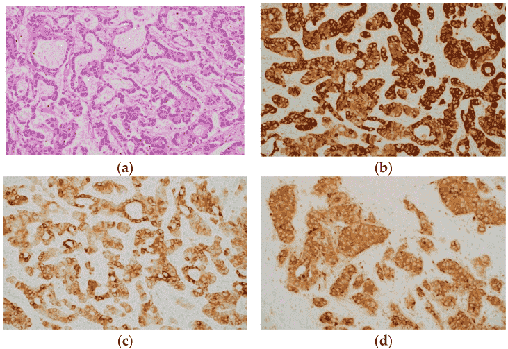 Fig.1 Histological appearance (a) and immunohistochemical staining (b–d) of intrahepatic cholangiocarcinoma (ICC).1
As a reputable global supplier, Creative Biolabs is committed to providing customers with our customized natural autoantibodies (NAA) services and optimized IHC solutions to accelerate the progress of your projects. Please contact us for more details.
Reference
- Takahashi, Yoshihisa, et al. "Application of immunohistochemistry in the pathological diagnosis of liver tumors." International Journal of Molecular Sciences 22.11 (2021): 5780.
Related Services:
- Enzyme-Linked Immunosorbent Assay (ELISA)
- Western Blot
- Cryogenic Electron Microscopy (Cryo-EM)
- Site-Directed Mutagenesis Mapping