

NAA Services for Anti-Myofibrillar Proteins
Creative Biolabs is an undisputed global leader in the natural autoantibody (NAA) and diseases diagnosis areas. Aided by innovative and diversified platforms, Creative Biolabs provides comprehensive services for various biomarkers against a wide variety of diseases. Having accumulated abundant experience in the successful completion of many NAA-related projects, we now offer a full range of anti-myofibrillar proteins services for diseases diagnosis and therapeutic monitoring.
Background of Myofibrillar Proteins
Myofibrillar proteins (MPs), comprising approximately 50% of total muscle proteins, are generally considered to be insoluble in solutions of low ionic strength (< 0.2 M), requiring high concentrations of salt (> 0.3 M) for solubilization. MPs exist as multiple isoforms that derive from multigene families and MP isogenes are differentially expressed in various muscle types and fiber types but can be co-expressed within the same fiber. The variable expression of MP isoforms is a major determinant of the contractile properties of skeletal muscle fibers. In early myoblasts, MPs form stable pre-myofibrillar assemblies surrounding the nucleus and raise the possibility that these initial assemblies may play an organizing role during subsequent early stages of myofibrillogenesis. Antibodies against various myofibrillar proteins were measured by ELISA in sera of 31 myasthenic patients, and finally, antibodies against myosin, tropomyosin, troponin, alpha-actinin, and actin were detected respectively.
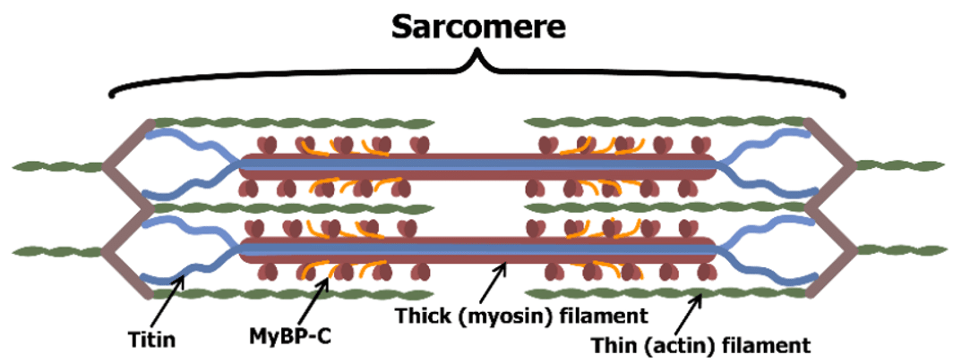 Fig.1 The structure of muscle sarcomere with thick and thin filaments.1
The Role of Anti-myofibrillar Proteins Antibody in Myasthenia Gravis (MG)
There is a substantial body of information to indicate alterations in MPs including actin, myosin, tropomyosin, troponin, titin, desmin, and myosin-binding protein C in conditions such as hyperthyroidism, hypothyroidism, myasthenia gravis (MG), and diabetes. Studies reported a strong association between the occurrence of anti-skeletal muscle (SM) antibodies and the presence of thymoma in patients with MG. Antibodies to MPs (titin, myosin, actin, actomyosin) are found in 85% of patients with thymoma and maybe the first evidence of thymoma in some cases. For example, anti-titin antibodies are a sensitive marker of thymoma associated with MG (MG-T) in patients 60 years and younger.
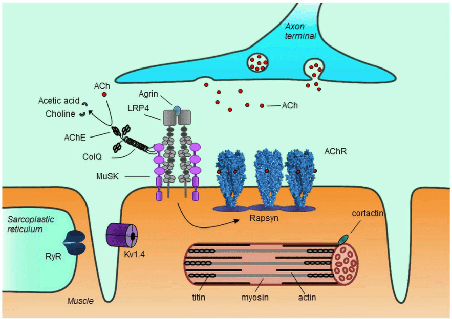 Fig.2 Role of myofibrillar proteins in MG.2
What We Can Do about NAA?
Based on our well-established platforms and experienced scientists, we can provide comprehensive NAA services to cover every step of your program, from NAA detection, NAA profiling, to NAA epitope mapping. A wide spectrum of NAA products is available for your choice.
Creative Biolabs provides a full range of high-affinity anti-myofibrillar proteins autoantibody analysis services for the diagnosis of MG. Our high-quality services will contribute greatly to the success of your project. If you are interested in the service we provide, please feel free to contact us for more detail and information.
References
- Bobyleva, Liya G., et al. "Myosin binding protein-C forms amyloid-like aggregates in vitro." International Journal of Molecular Sciences 22.2 (2021): 731.
- Lazaridis, Konstantinos, and Socrates J. Tzartos. "Autoantibody specificities in myasthenia gravis; implications for improved diagnostics and therapeutics." Frontiers in immunology 11 (2020): 212.
Related Services:
- NAA Services for Anti-Acetylcholine Receptor (AChR)
- NAA Services for Anti-Muscle-specific Tyrosine Kinase (MuSK)
- NAA Services for Anti-Lipoprotein-related Protein Receptor 4 (LRP4)
- NAA Services for Anti-Ryanodine Receptor (RyR)
- NAA Services for Anti-Titin Antibody
- NAA Services for Anti-Interferon-α and Interleukin
- NAA Services for Anti-VGKC (Kv1.4)