

NAA Services for Anti-Ryanodine Receptor (RyR)
Equipped with extensive experience, advanced platform, and professional technical team, Creative Biolabs provides a comprehensive set of natural autoantibody (NAA) analysis services for the biomarker ryanodine receptor (RyR) to push the diagnosis of myasthenia gravis (MG). With years of experience in the field of disease diagnosis, our high-quality services will contribute greatly to the success of your project.
Background of Anti-ryanodine Receptor (RyR) Antibody
Ryanodine receptor (RyR), a Ca2+ release channel of the sarcoplasmic reticulum, is essential for the excitation-contraction coupling in striated muscle. Anti-RyR antibodies recognize a region near the N-terminus on the RyR, which seems to be of importance for RyR regulation. These antibodies cause allosteric inhibition of RyR function in vitro, inhibiting Ca2+ release from the sarcoplasmic reticulum in vitro. Anti-RyR antibodies are mainly expressed in the IgG1 and IgG3 subclasses and may have pathogenetic relevance in thymoma-associated myasthenia gravis (MG-T).
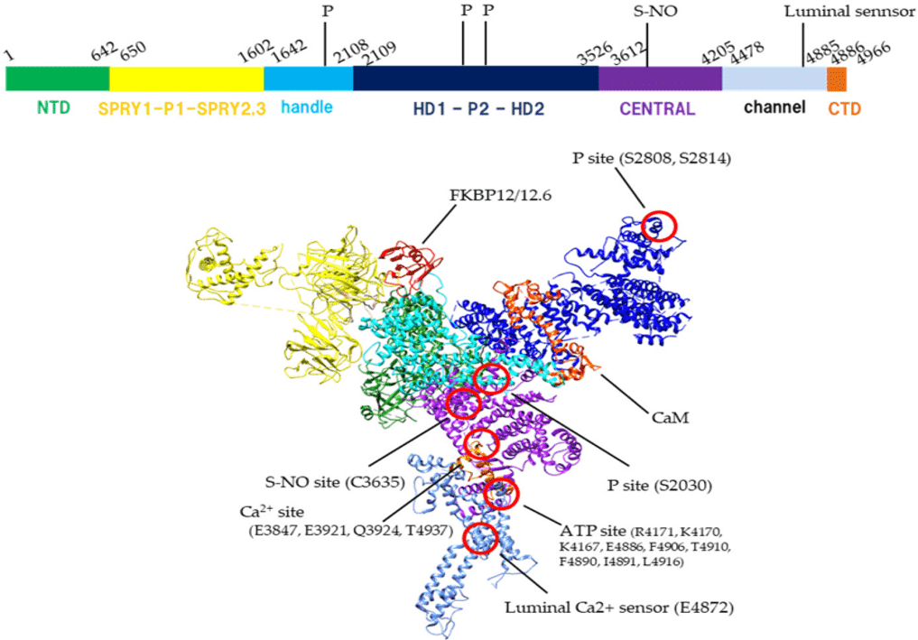 Fig.1 Structure of RyR.1
The Role of Anti-RyR Antibody in Myasthenia Gravis (MG)
MG is an organ-specific autoimmune disorder. Some MG patients have autoantibodies against skeletal muscle antigens in addition to the acetylcholine receptor (AChR) and muscle-specific receptor tyrosine kinase (MuSK). Two major antigens for non-AchR antibodies in MG are the RyR and titin. Anti-RyR antibodies are found mainly in thymoma MG patients and in a few late-onset MG patients and correlate with a severe MG disease. RyR antibody levels correlated positively with the severity of MG. Complement-activating RyR autoantibodies occurred with higher frequency in sera of thymoma MG than of late-onset MG. RyR IgG 1 antibodies occurred more often in severe MG than in mild and moderate disease groups. Mean total IgG and IgG 1 titin and RyR antibody titers fell during long-time patient observation together with an improvement of the MG symptoms.
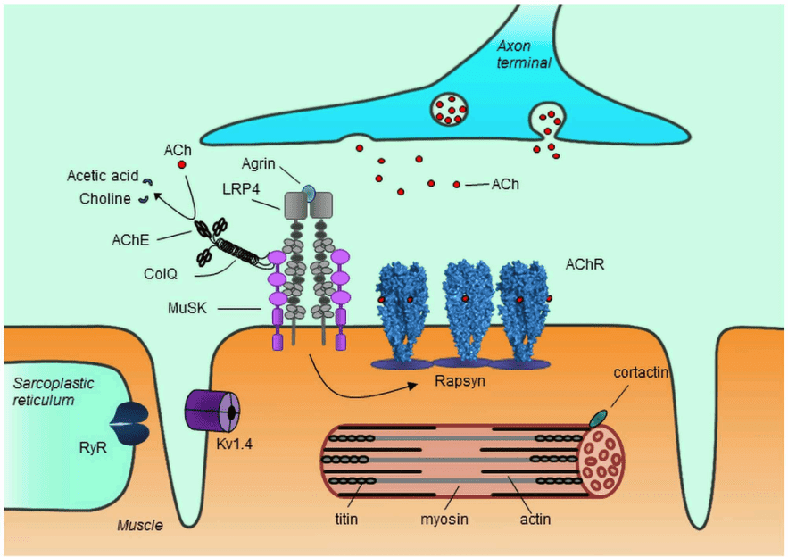 Fig.2 Role of auto-antigens of MG at the neuromuscular junction and in muscle.2
What We Can Do about NAA?
Equipped with our well-established platforms and experienced scientists, we can provide comprehensive NAA services, from NAA detection, NAA profiling, to NAA epitope mapping. A wide spectrum of NAA products is available for your choice.
Features of Our Services
- High-quality NAA service without large scale repeats
- Professional technical team and advanced platform to save your cost and valuable time
- Across-the-board technical guidance
- Fast turnaround time to ensure your program schedule
- Best after-sale service
Studies illustrated that monitoring the titer of special autoantibody can be useful for disease early diagnosis and status assessment as well as decision making during treatment. Therefore, it is necessary to develop more effective and convenient methods for the screening and early diagnosis of MG. As a well-recognized leader in disease diagnosis and with over a decade of experience, innovative and diversified NAA platforms, Creative Biolabs is dedicated for developing serum NAA tests against RyR biomarker and offering you a comprehensive set of high-quality NAA analysis services. We also provide custom services based on the requirements of the clients to meet the specific demand. Please don’t hesitate to contact us for more information.
References
- Kobayashi, Takuya, Nagomi Kurebayashi, and Takashi Murayama. "The ryanodine receptor as a sensor for intracellular environments in muscles." International Journal of Molecular Sciences 22.19 (2021): 10795.
- Lazaridis, Konstantinos, and Socrates J. Tzartos. "Autoantibody specificities in myasthenia gravis; implications for improved diagnostics and therapeutics." Frontiers in immunology 11 (2020): 212.
Related Services:
- NAA Services for Anti-Acetylcholine Receptor (AChR)
- NAA Services for Anti-Muscle-specific Tyrosine Kinase (MuSK)
- NAA Services for Anti-Lipoprotein-related Protein Receptor 4 (LRP4)
- NAA Services for Anti-Titin Antibody
- NAA Services for Anti-Interferon-α and Interleukin
- NAA Services for Anti-VGKC (Kv1.4)
- NAA Services for Anti-Myofibrillar Proteins