Bispecific antibodies (BsAbs) are engineered antibody-derived proteins with two or more distinct binding specificities within one molecule, mainly produced by quadroma technology, chemical conjugation, or genetic approaches. A vast array of formats of BsAbs have been generated by different technologies to cater to research and clinical uses, including applications in diagnostics.
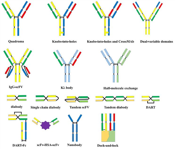
Fig. 1 Molecular formats of BsAbs.1
Owing to the potential of concurrent detection of multiple antigens or combining assay markers with antigen-binding sites, along with the flexibility and convenience in design, BsAbs have been considered as promising diagnostic immunoprobes with several remarkable advantages.
BsAbs have been widely used for the detection of viral or bacterial infectious diseases and in cancer diagnosis. They can participate in pre-targeting strategies in clinical diagnosis and provide better imaging for the early detection and diagnosis of tumors.
For instance, BsAbs that target human red blood cells (RBCs) and hepatitis B virus surface antigen (HBsAg) show encouraging diagnostic performance with high sensitivity (97.7%) and specificity (100%) in the detection of HBsAg in blood samples without special equipment or training. Furthermore, it is demonstrated that BsAbs targeting Lipoarabinomannan (LAM) and horseradish peroxidase (HRPO) significantly reduce the time required for detection of tuberculosis bacteria, from 2-6 weeks (traditional laboratory culture) to less than 2h, also ensuring high specificity (100%) and sensitivity (64%).²
In medical imaging for cancer and potential lesions, pre-targeted detection based on BsAbs and small radiolabeled molecules exhibits high binding specificity, sensitivity, and safety, which compensates for the limitations of 18F fluorodeoxyglucose positron emission tomography (FDG-PET) in recognizing small tumors (less than 1 cm) and inflammatory or infectious lesions.
Paper Title | Promising clinical performance of pretargeted immuno-PET with anti-CEA bispecific antibody and gallium-68-labeled IMP-288 peptide for imaging colorectal cancer metastases: a pilot study |
Journal | European Journal of Nuclear Medicine and Molecular Imaging |
Published | 2021 |
Abstract | Colorectal carcinoma (CRC) is a common cancer that takes more than 500,000 lives per year. To optimize the existing methods of Imaging workup, immuno-PET methods, using a radiolabelled small molecule and a nonradioactive BsAb that binds to both tumor antigen and the specific small molecule to localize the tumor, has been proposed as a strategy. The BsAb TF2 is designed to simultaneously identify CEA, a representative target for CRC in immuno-PET), and histamine succinate glycine (HSG) motif. The study aims to assess the diagnostic performance of the immuno-PET method in comparison with conventional imaging and FDG-PET in patients with metastatic CRC, using TF2 combined with a 68Ga -labeled HSG peptide, 68Ga-IMP288. The result reveals that pretargeted immuno-PET using TF2/68Ga -IMP288 has promising potential in imaging workup, as well as sensitivity and safety. |
Result |
For a per-patient analysis, positive results identified by immuno-PET were observed in 9/10 patients. One patient (patient 5) was considered false negative by immuno-PET, whose unique lung metastasis was recognized by FDG-PET and histology. For per-lesion analysis, immuno-PET was positive in 12/14 lesions. Both FDG-PET and conventional imaging failed to identify multiple liver metastases in patient 2, which was only visualized by immuno-PET and confirmed by histology. For mediastinal sarcoidosis nodes in patient 10 and a chronic anastomotic fistula of the rectum in patient 11, immuno-PET was negative while FDG-PET was positive. The specificity, sensitivity, positive predictive value, and negative predictive value were 88%, 100%, 100%, and 67% for immuno-PET, significantly higher than those of other methods in this study (76%, 67%, 87%, and 33% for FDG-PET; 25%, 82%, 82% and 25% for the combination of EUS/CT/ MRI). Median SUVmax, MTV, and TLG respectively for immuno-PET lesions were 7.65 [3.98-13.94, SD 3.37], 8.63 cm³ [1.98-46.64; SD 14.83], and 37.90 cm³ [8.07-127.5; SD 43.47].
Fig. 2 Imaging workup of multiple liver metastases only visualized by immuno-PET.3 |
With our high-caliber BsAbs development platforms, Creative Biolabs is dedicated to providing customers with qualified and reliable one-stop services related to BsAbs, from design, engineering, purification to analysis, and manufacturing. We are here to assist your research for both scientific and clinical purposes. Please do not hesitate to contact us for more detailed inquiries.
References
1. Fan, Gaowei, et al. "Bispecific antibodies and their applications." Journal of hematology & oncology 8 (2015): 1-14.
2. Byrne, Hannah, et al. "A tale of two specificities: bispecific antibodies for therapeutic and diagnostic applications." Trends in biotechnology 31.11 (2013): 621-632.
3. Touchefeu, Y., et al. "Promising clinical performance of pretargeted immuno-PET with anti-CEA bispecific antibody and gallium-68-labeled IMP-288 peptide for imaging colorectal cancer metastases: a pilot study." European Journal of Nuclear Medicine and Molecular Imaging 48 (2021): 874-882.
Welcome! For price inquiries, we will get back to you as soon as possible.
INQUIRYSERVICES
PRODUCTS
PLATFORMS