Exosome Fluorescent Labeling Service
Overview Services Features FAQs
Fluorescent labeling involves attaching fluorescent dyes or proteins to exosomes, allowing researchers to track their movement and interactions. Creative Biolabs provides excellent exosome fluorescent labeling services, enabling the study of exosome uptake by recipient cells, their distribution within tissues, and their clearance from the body.
Fluorescent Labeling
Fluorescent labeling is a technique used in molecular biology, biochemistry, and related fields to visualize and track molecules or structures of interest. It involves attaching fluorescent molecules, called fluorophores, to specific target molecules. Fluorescent labeling has revolutionized many areas of biological research by providing a powerful tool for studying complex biological processes at the molecular level with high sensitivity and specificity.
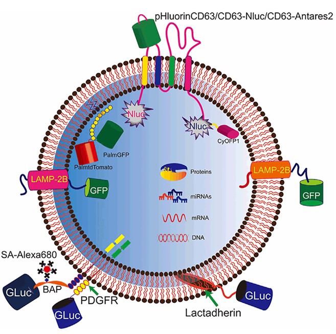 Fig. 1 Genetic engineering for EV labeling.1
The biological characteristics of exosomes can be investigated with great effectiveness thanks to fluorescent labeling. For example, total exosomes can be labeled by fluorescent dyes, which can be used to detect the distribution and location as well as trafficking of exosomes. Besides, antibodies labeled by fluorescent dyes have a high affinity with specific antigens on the exosomes. Hence, antibody fluorescent labeling allows us to monitor characteristics of specific exosomes in vivo.
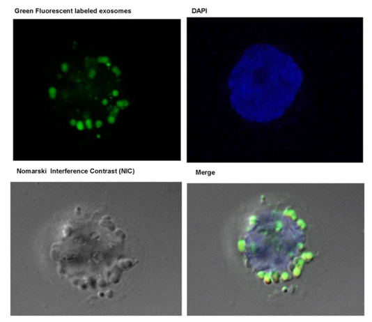 Fig. 2 Confocal microscopy- Green Fluorescent labeled exosomes were taken up by THP1 monocytes as early as 6 h after Treatment.2
Exosome Fluorescent Labeling Service
-
Identification of Exosome Characteristics—Fluorescence conjugated antibody allows to visualize and recognize individual exosomes by a confocal laser scanning microscope or flow cytometry. Our exosome fluorescent labeling platform helps identify the distribution and concentration of the interested exosomes, making it a promising source for diagnostics.
-
Tracking Exosome in Vivo—exosome can be used as a vector for drug delivery to target tissue. Our fluorescence labeling service makes it possible to visualize and monitor the movement of exosomes bearing drugs in vivo, providing an effective tool for the optimization of treatment protocols in time.
Advantages and Features of Exosome Fluorescent Labeling Service in Creative Biolabs
-
The highest sensitivity and specificity
-
Minimal impact on exosome characteristics
-
An extensive degree of adaptability in the dye labeling procedure
-
All-around technical service
FAQs
Q: Why need exosome fluorescent labeling service?
A: Exosome fluorescent labeling service is commonly used in research studies to track exosomes in vitro and in vivo, allowing for the study of their release, uptake, and distribution. The function of exosomes in several biological processes and disease states can be better understood with the help of the service.
Q: What types of fluorescent labels can be used for exosome labeling?
A: Common fluorescent labels used for exosome labeling include fluorescent dyes such as PKH26, PKH67, DiI, and DiO, as well as fluorescent proteins like GFP and RFP. These labels emit fluorescence when excited by specific wavelengths of light, enabling their visualization using fluorescence microscopy or flow cytometry.
Q: How is the exosome fluorescent labeling service performed?
A: Exosome fluorescent labeling service typically involves incubating exosomes with the chosen fluorescent label, allowing for the dye or tag to bind to the exosome membrane. The labeled exosomes are then purified and characterized before being returned to the customer for further analysis.
Q: What are the benefits of exosome fluorescent labeling service?
A: Exosome fluorescent labeling service allows for the visualization and tracking of exosomes in biological samples, enabling researchers to gain valuable insights into exosome biology and function.
Please contact us with Creative Biolabs' exosome scientist team to learn more about what we can offer for your exosome tracing project.
References
-
Liu, Qisong, et al. "Tracking tools of extracellular vesicles for biomedical research." Frontiers in Bioengineering and Biotechnology 10 (2022): 943712. Under Open Access license CC BY 4.0, without modification.
-
Momen-Heravi, Fatemeh, et al. "Exosomes derived from alcohol-treated hepatocytes horizontally transfer liver specific miRNA-122 and sensitize monocytes to LPS." Scientific reports 5.1 (2015): 9991. Under Open Access license CC BY 4.0, without modification.
For Research Use Only. Cannot be used by patients.
Related Services: