Tissue Exosome Research & Applications
Introduction Advantages Applications Services FAQs
The Untapped Potential of Tissue Exosomes
Exosomes are nanoscale exosomes (30–150 nm) secreted by nearly all cell types and present in biofluids like plasma and urine, as well as in tissue interstitial spaces. Far from cellular debris, exosomes are active biological messengers that transfer proteins, lipids, nucleic acids (e.g., miRNA, mRNA, lncRNA), and metabolites between cells, influencing processes like proliferation, differentiation, and apoptosis.
While most studies focus on exosomes from biofluids, tissue-derived exosomes—produced locally within tissues—offer a more accurate snapshot of the pathological microenvironment. Unlike circulating exosomes, which can be diluted by systemic signals, tissue exosomes provide a clearer, more specific view of localized disease states, making them particularly valuable for conditions like solid tumors or organ-specific injuries.
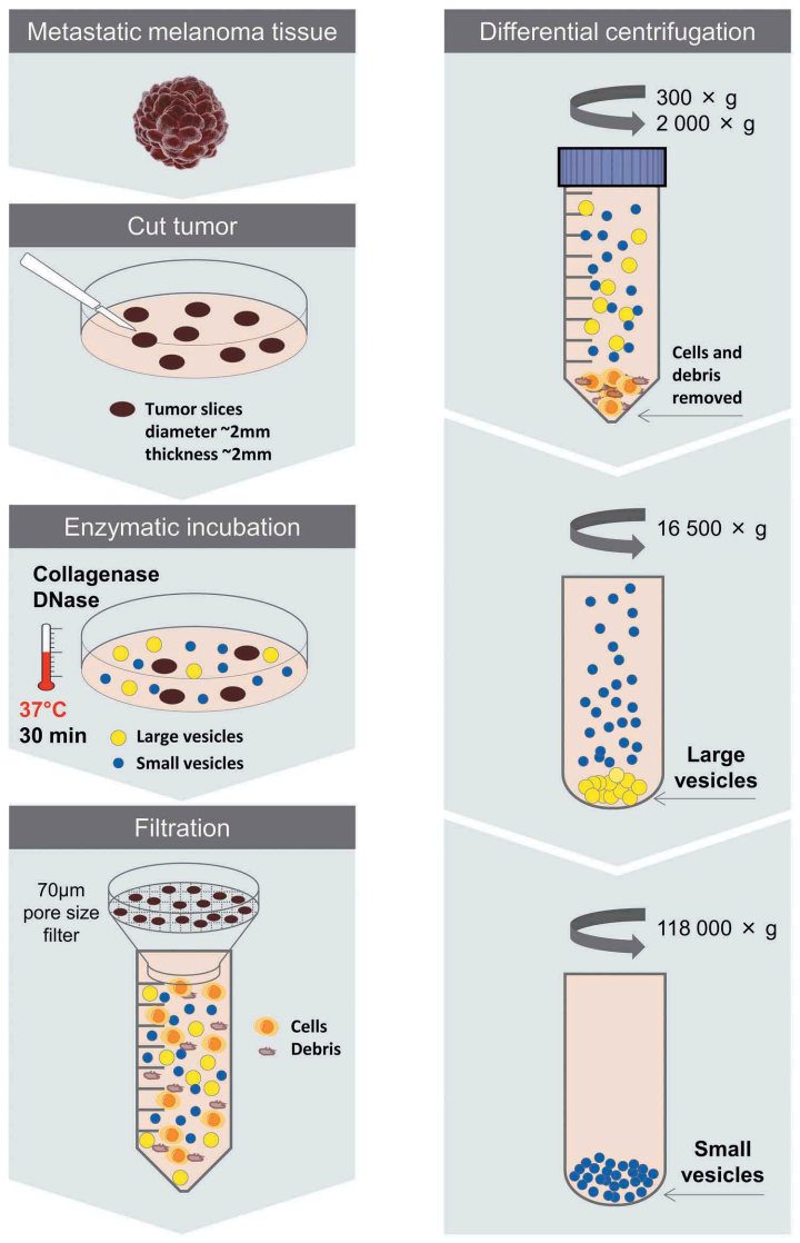 Fig.1 Schematic overview of the centrifugation-based protocol used to isolate vesicles from metastatic melanoma tissues.1
This shift in focus marks a new era in biomarker discovery and targeted therapy. Tissue exosomes allow for earlier and more precise diagnosis and offer therapeutic potential beyond drug delivery. By influencing and reprogramming recipient cells, exosomes can actively direct biological responses such as immune modulation, tissue repair, and tumor suppression—paving the way for next-generation, cell-instructive therapies. Creative Biolabs has established a comprehensive one-stop solution for tissue-derived exosome research and looks forward to collaborating with you to advance this exciting field.
Advantages: Why Tissue Exosomes Stand Apart
Compared with exosomes derived from body fluids, tissue exosomes possess distinct advantages that position them as superior tools for advanced disease research and therapeutic development.
Table 1. Comparative Advantages: Tissue Exosomes vs. Biofluid Exosomes
|
Feature
|
Tissue Exosomes
|
Biofluid Exosomes (e.g., Plasma, Serum)
|
|
Origin & Specificity
|
Predominantly from cells within the local tissue interstitial space
|
Mixed population from various tissues and organs throughout the body
|
|
Authenticity
|
High; directly reflects local pathological changes
|
Lower; broader, systemic reflection, potential for confounding signals
|
|
Exclusivity
|
High; specific to the local microenvironment
|
Lower; less specific, reflects overall physiological state
|
|
Biomarker Utility
|
Ideal for precise, localized disease markers
|
Useful for systemic markers, but less specific for tissue-level changes
|
|
Complexity of Analysis
|
Reduced complexity compared to whole bodily fluids
|
High complexity due to diverse cellular origins and contaminants
|
Applications: Pioneering Disease Research with Tissue Exosomes
Tissue exosomes are not merely a research curiosity but a powerful platform with immense potential across diagnostics and therapeutics. Their cargo, including specific proteins, miRNAs, and lncRNAs, can serve as indicators for early detection, disease staging, and monitoring therapeutic response. The utility of tissue exosomes is further amplified by their tissue-specific cargo, offering tailored insights into various diseases. The currently reported studies on tissue exosomes are mainly divided into the following aspects.
A Comprehensive, End-to-End Service Portfolio
Creative Biolabs offers a comprehensive, end-to-end service portfolio designed to support all aspects of tissue exosome research. Our extensive experience in molecular and cell biology allows us to provide a holistic understanding of tissue exosome functions, from initial isolation and characterization to advanced cargo profiling and engineering.
-
Creative Biolabs uses a self-developed tissue processing method to successfully extract exosomes from complex tissues.
-
Obtain the expression profile of exosomes in the microenvironment of diseased tissues in vivo.
Creative Biolabs offers
exosomal RNA profiling services, such as
exosomal miRNA sequencing,
exosomal mRNA sequencing, and
exosomal lncRNA sequencing, to help analyze gene expression profiles for disease-related tissue exosomes.
-
Combine with body fluid exosome expression profiles to screen out more specific disease diagnostic and prognostic markers.
Creative Biolabs provides
exosomal protein profiling services and
exosomal RNA profiling services, which allow researchers to compare tissue-derived exosomes with those found in body fluids, enhancing the accuracy of biomarker discovery.
-
Discover the signaling pathways involved in exosomes in the microenvironment of diseased tissues in vivo and provide possible targets for the disease treatment.
-
Combine with exosomes in the microenvironment of other tissues and discover the exosome communication mechanism of two different tissues in vivo.
Through our data analysis and comparison, we facilitate comprehensive studies on exosome communication across different tissue types, enabling a deeper understanding of how exosomes mediate inter-tissue communication in disease progression.
If you want to research tissue exosomes, please contact us.
FAQs
Q: How do tissue exosomes provide more specific insights compared to exosomes from biofluids like blood?
A: Tissue exosomes are predominantly secreted by cells within the local tissue's interstitial space, offering a direct and authentic representation of the communication network within a specific pathological microenvironment. Unlike exosomes found in systemic biofluids, which can be diluted or reflect signals from various sources, tissue exosomes capture real-time, localized pathological changes with unparalleled precision. For instance, proteomic analysis has revealed hundreds of differentially expressed proteins in cancer tissue-derived exosomes that are specific to the localized disease state, a level of detail often not attainable with circulating exosomes.
Q: What animal models are utilized for in vivo exosome functional studies?
A: A variety of animal models, including rodents and larger models when appropriate, are utilized to study exosome biodistribution and mechanistic pathways in vivo. Models are selected and customized based on the specific requirements of each research project.
Q: Can exosome labeling and in vivo study services be customized to fit specific research needs?
A: Yes, the technical team designs customized experimental plans in close consultation with clients, based on in-depth communication and demand analysis. Real-time adjustments and optimizations are also made during the experiment to provide the most valuable data.
Q: What is the typical turnaround time for results from exosomal proteomic detection service?
A: For ultra-sensitive targeted exosomal proteomic detection service, results are generally available within 8-12 weeks, depending on the complexity of the analysis and the specific panel requirements. Expedited services may be available upon discussion. For mass spectrometry-based exosome characterization, it typically takes 8-10 weeks from sample receipt to final report delivery.
Q: How is the dependability and accuracy of the outcomes guaranteed?
A: Cutting-edge technology coupled with strict quality control procedures is employed to ensure the precision and reliability of findings. Each sample undergoes multiple validation steps, and adherence to industry standards for proteomic analysis and exosome characterization is maintained. Experienced professionals and a whole-process technical trace service further ensure accurate experimental data.
Reference
-
Crescitelli, Rossella et al. "Subpopulations of exosomes from human metastatic melanoma tissue identified by quantitative proteomics after optimized isolation." Journal of exosomes vol. 9,1 1722433. 11 Feb. 2020, doi:10.1080/20013078.2020.1722433. Distributed under Open Access license CC BY 4.0, without modification.
For Research Use Only. Cannot be used by patients.
Related Services: