In Vitro Functional Services of Exosomes

For in vitro exosomal function exploration, exosome samples are usually labeled first, and the following researches will realize by tracking labeled exosomes. Creative Biolabs has established a comprehensive one-stop exosomal function in vitro research platform. Our services cover every step of the exploration, including the labeling and tracking, as well as the subsequent cell-based exosomal function researches.

Exosomes Labeling

In the published exosomes researches, most of the exosomes are labeled with lipophilic dyes, and they have many applications in vivo and in vitro. Lipophilic dyes are mainly divided into two categories. The first category is PKH67 (green fluorescence)/PKH26 (red fluorescence). Because they can stably bind to the lipid bilayer membrane of exosomes, and have better dyeing effects as well as the application. widely. The second category is the lipophilic dyes of the Di series, including DiI (orange fluorescence), DiO (green fluorescence), DiD (red fluorescence), and DiR (dark red fluorescence). Among them, the infrared fluorescence of DiR can penetrate cells and tissues and is used for tracking in in vivo imaging. In addition to lipophilic dyes that have been widely used, penetrant dyes, including carboxyfluorescein diacetate succinimidyl ester (CFSE) and calcein acetyl methyl ester (Calcein-AM), can penetrate exosomal membranes into the exosomes. CFSE and Calcein-AM can not only label exosomes but also verify the integrity of exosomes.

In this method, exosome-specific membrane proteins (such as CD9, CD63, and CD81) and fluorescent proteins are constructed as the expression elements into plasmids. Constructed plasmids are packaged into lentiviruses, and then the cells are infected with the lentiviruses so that the exosomes secreted by these cells are labeled by fluorescent proteins.

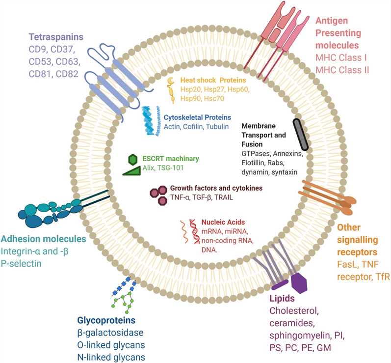
Different exosomal membrane proteins are involved in the fusion of targeting/labeling moieties for surface functionalization. Fig.1 Different exosomal membrane proteins are involved in the fusion of targeting/labeling moieties for surface functionalization. (Salunkhe, 2020)

Add the labeled exosomes to the recipient cell culture medium and co-culture with the recipient cells to observe cell function changes, such as cell proliferation, migration, and invasion, cell apoptosis, etc.

In Vitro Function of Exosomes

Services at Creative Biolabs

Creative Biolabs has rich experience in exosomal function researches and has established a comprehensive technology platform that covers the whole process of exosome function study. Our platform offers a variety of exosomal function research services including but not limited to:

  • Exosomes labeling or tracing
  • NTA analysis of mass production size and quantity of exosomes
  • Stability test of electroporation exosomes
  • Exosomes uptake experiment
  • CCK8 cell proliferation experiment
  • Flow Cytometry Test of Apoptosis
  • Flow cytometry cell cycle
  • Cell Migration Transwell
  • Cell invasion test

If you are interested in exosomal function in vitro research, or you have any other questions about our services, please don't hesitate to contact us for more information.

Reference

  1. Salunkhe, S.; Dheeraj.; et al. Surface functionalization of exosomes for target-specific delivery and in vivo imaging & tracking: Strategies and significance. Journal of Controlled Release. 2020. 326:599-614.
For Research Use Only. Cannot be used by patients.
Related Services:
Online Inquiry
Get resources and offers direct to your inbox: Submit
Inquiry Basket