Creative Biolabs is offering the most comprehensive services for antibody development projects. With strict regulation and effective execution, we are dedicated to providing the most valuable solutions to complete your projects.
HighlightsWith over a decade of experience in phage display technology, Creative Biolabs can provide a series of antibody or peptide libraries that are available for licensing or direct screening. These ready-to-use libraries are invaluable resources for isolating target-specific binders for various research, diagnostic or therapeutic applications. Highlights
Creative Biolabs has established a broad range of platforms for developing novel antibodies or equivalents. These cutting-edge technologies enable our scientists to meet your demands from different aspects and tailor the most appropriate solution that contributes to the success of your projects.
HighlightsWith deep understanding in antibody-related realms and extensive project experience, Creative Biolabs offers a variety of references to help you learn more about our capacities and achievements, including infographic, flyer, case study, peer-reviewed publications, and all kinds of knowledge that can assist your projects. You are also welcome to contact us directly for more specific solutions.
HighlightsGet a real taste of Creative Biolabs, one of the most professional custom service providers in the world. We are committed to providing highly customized comprehensive solutions with the best quality to advance your projects.
HighlightsApoptosis plays a pivotal role in maintaining the balance and health of multicellular organisms by regulating the life and death of individual cells. This article will delve deep into the world of apoptosis, exploring its significance, mechanisms, and the role it plays in various aspects of biology.
Apoptosis, often referred to as programmed cell death, is a fundamental biological process that plays a critical role in maintaining the health and functionality of multicellular organisms. It is a precisely regulated mechanism by which cells undergo self-destruction in a controlled and orchestrated manner. Apoptosis is essential for the normal development, growth, and maintenance of tissues, as well as for the proper functioning of the immune system.
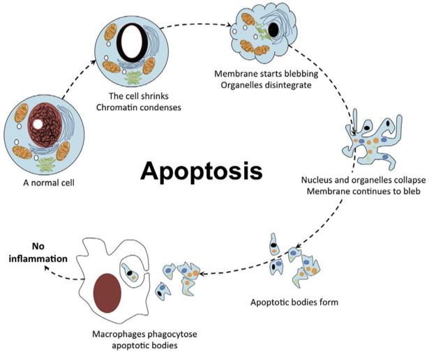 Fig 1. Cytology of apoptosis.1
Apoptosis is characterized by a series of distinct and well-defined features that set it apart from other forms of cell death:
During apoptosis, a cell undergoes a process of shrinkage, leading to a reduction in cell volume. This is in stark contrast to necrosis, another form of cell death, where cells swell and eventually burst.
One of the early hallmarks of apoptosis is the condensation of chromatin within the cell nucleus. This results in the formation of dense, compact structures visible under a microscope.
Perhaps one of the most distinctive features of apoptosis is the fragmentation of DNA. As the cell progresses through apoptosis, its DNA is cleaved into smaller fragments, producing a characteristic ladder-like pattern when analyzed by gel electrophoresis.
Apoptotic bodies are small membrane-bound vesicles that contain cellular components and organelles. These bodies are generated during the final stages of apoptosis and are subsequently engulfed and digested by neighboring cells or phagocytes.
Unlike necrosis, which often triggers inflammation due to the release of cellular contents into the surrounding tissue, apoptosis typically occurs without provoking an inflammatory response. This is crucial for maintaining tissue integrity.
Apoptosis is a highly regulated process controlled by a network of molecular pathways. Two primary pathways, the intrinsic (mitochondrial) and extrinsic (death receptor) pathways, converge to activate a group of enzymes known as caspases. These enzymes play a central role in executing the various stages of apoptosis:
 Fig 2. Mechanisms of apoptosis.2
Initiated from within the cell, the intrinsic pathway is often triggered by internal signals, such as DNA damage or the presence of abnormal proteins. The mitochondria play a pivotal role in this pathway. When cells receive signals to undergo apoptosis, the mitochondria release certain proteins, including cytochrome c, into the cytoplasm. Cytochrome c then activates caspases, initiating the apoptotic cascade.
In contrast, the extrinsic pathway is activated by external signals. These signals are typically received by death receptors on the cell surface. When specific molecules, such as tumor necrosis factor (TNF) or Fas ligand, bind to these death receptors, they trigger the activation of caspases and the initiation of apoptosis. This pathway is particularly important in the immune system's response to infected or damaged cells.
The regulation of apoptosis is a complex and finely tuned process that involves a balance between pro-apoptotic and anti-apoptotic factors. Key regulatory elements include:
The Bcl-2 family of proteins plays a central role in regulating apoptosis. Anti-apoptotic members, such as Bcl-2 and Bcl-xL, promote cell survival by preventing the release of cytochrome c from the mitochondria. Pro-apoptotic members, like Bax and Bak, promote cytochrome c release and apoptosis. The balance between these opposing forces dictates whether a cell lives or dies. (Learn more about our Magic™ membrane protein production services.)
IAPs are proteins that can inhibit caspases, preventing their activation and blocking apoptosis. Overexpression of IAPs is often observed in cancer cells, contributing to their resistance to cell death. Strategies aimed at targeting IAPs have been explored in cancer therapy.
The tumor suppressor protein p53 is a critical regulator of apoptosis, especially in response to DNA damage. When DNA damage occurs, p53 can activate the expression of pro-apoptotic proteins, promoting cell death to prevent the propagation of damaged DNA. Additionally, p53 can suppress anti-apoptotic genes, reinforcing the apoptotic response.
Cells can receive signals from their environment, known as survival factors, which promote cell survival by inhibiting apoptosis. These factors are essential for the survival of specific cell types, such as neurons and immune cells. For example, nerve growth factor (NGF) acts as a survival factor for neurons, preventing them from undergoing apoptosis and supporting their growth and function.
Dysregulation of apoptosis can have profound implications for health and disease, impacting various physiological processes and contributing to the development and progression of numerous medical conditions.
Cancer is often characterized by the evasion of apoptosis. Cancer cells can acquire mutations that disrupt the normal apoptotic pathways, allowing them to survive and proliferate uncontrollably. Understanding the mechanisms of apoptosis has led to the development of targeted therapies that aim to induce apoptosis in cancer cells. For instance, some chemotherapeutic drugs work by promoting apoptosis in rapidly dividing cancer cells. Additionally, targeted therapies specifically designed to inhibit anti-apoptotic proteins like Bcl-2 are showing promise in cancer treatment.
In neurodegenerative diseases like Alzheimer's and Parkinson's, the death of neurons is a hallmark. Dysregulation of apoptosis has been implicated in these diseases, and researchers are exploring ways to modulate apoptosis to potentially slow disease progression. For example, drugs that target apoptosis-related pathways may help protect neurons from degeneration in Alzheimer's disease.
Autoimmune disorders can result from a failure of immune cells to regulate apoptosis properly. In some cases, immune cells may mistakenly attack healthy tissues due to a lack of apoptosis regulation. Understanding these defects in apoptosis can provide insights into the development of treatments that restore proper immune regulation in autoimmune diseases.
Aging is associated with a decline in the regulation of apoptosis. Accumulation of cells resistant to apoptosis can contribute to tissue dysfunction and age-related diseases. Researchers are investigating strategies to rejuvenate aged tissues by restoring the balance between cell survival and cell death.
References
All listed services and products are For Research Use Only. Do Not use in any diagnostic or therapeutic applications.