Extracellular Vesicle Labeling Service
Overview Services Features FAQs
Overview
Why Label Extracellular Vesicles
Extracellular vesicles (EVs) can participate in the regulation of many important physiological and pathological processes by mediating intercellular communication. In-depth study of the biological behavior and function of EV and their correlation with major diseases will not only help reveal the pathogenesis of related diseases, but also provide new liquid biopsy markers for early diagnosis and prognosis evaluation of diseases. In addition, based on the natural delivery properties and good biosafety of EV, there has been an increasing amount of research on the use of EV to construct drug delivery systems in recent years. Therefore, in order to study the distribution, pharmacokinetics, biodegradation, in vivo stability, cell uptake, and the transport mechanism of loaded drugs in the circulation and cells, EV labeling and in vivo or in vitro tracing of EV are very critical.
Based on the actual needs of customers, Creative Biolabs has been committed to providing the most suitable EV labeling solution using molecules with fluorescent properties.
Services
Endogenous Labeling Service for Extracellular Vesicle
The endogenous labeling service of EV we provide is to fuse fluorescent proteins with marker proteins on the EV membrane by using genetic engineering means. For example, the expression elements of CD63 (marker protein on the EV membrane) and mCherry (red fluorescent protein) are constructed into a plasmid. This plasmid is then transfected into parental cells, causing the cells to secrete EVs with red fluorescent. The biggest advantage of this labeling method is that it can track the movement of EVs from parental cells to target cells. Dynamic monitoring of fluorescent proteins by fluorescence microscopy allows direct visualization of EV transfer in live culture and in vivo.
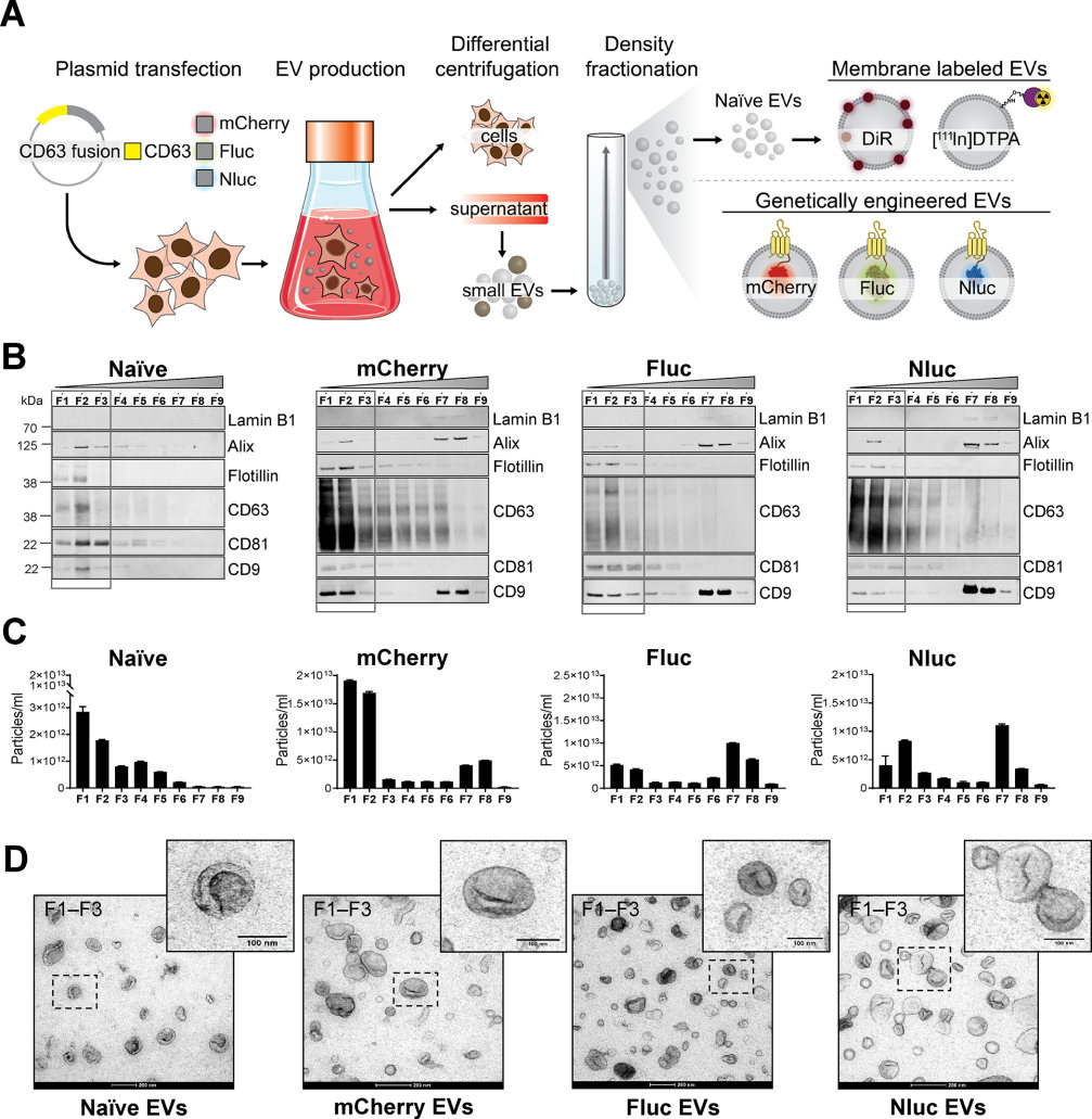 Fig.1 The production process of the membrane-labeled and engineered EVs.1,2
Exogenous Labeling Service for Extracellular Vesicle
The exogenous labeling service of EV we provide is by directly labeling the isolated EVs with lipophilic fluorescent membrane dyes. The basic principle is that this fluorescent dye can emit a strong fluorescent signal after inserting the aliphatic long tail into the lipid bilayer of EV. Common EV dyes include PKH67 (λex/λem = 490/502 nm), PKH26 (λex/λem = 551/567 nm), DiO (λex/λem = 484/501 nm), DiI (λex/λem = 549/565 nm ), DiR (λex/λem= 750/780 nm). These dyes can emit stable and persistent fluorescent signals on the EV membrane, which facilitates the observation of the interaction between EVs and target cells, especially in vitro.
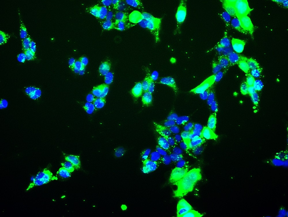 Fig.2 Extracellular vesicles are taken up by receptor cells. Green: DiO-labeled extracellular vesicles; Blue: the nuclei of receptor cells.
Features
-
Dual Labeling Strategies
-
Enhanced Visualization and Tracking
-
Minimal Impact on Vesicle Functionality
-
Comprehensive Support from Our Experienced Team
Creative Biolabs is a supplier that focuses on the research progress of EV and provides the most comprehensive EV research service. If you want to observe the trajectory and distribution of EV in your research, please contact us with your specific needs. Our professional team will recommend the most suitable labeling service based on the source of EVs, separation method, and experimental purpose to help you observe EV more accurately. Not only that, at Creative Biolabs, you can also complete EV isolation and Exosome Characterization, EV profiling, engineered EV construction, EV functional research and verification, and EV large-scale isolation and purification.
FAQs
Q:How do I choose between endogenous and exogenous labeling for my study?
A: The choice of labeling method depends on your downstream experiments and research goals, whether endogenous or exogenous labeling. Endogenous labeling, achieved through donor cell engineering, is ideal for long-term studies where stable and consistent labeling of extracellular vesicles is required. Exogenous labeling, using dyes, is more suitable for short-term studies or when rapid and flexible labeling is needed. If you are unsure which labeling method to choose, our technical team will recommend the best strategy based on our communication with you.
Q: What detailed information should I provide in order to develop an exosome labeling plan?
A: When designing a labeling experiment, please provide details on the type of labeling desired (endogenous or exogenous), the intended use of the labeled vesicles (e.g., in vitro or in vivo studies), and the target cell types or tissues any specific requirements.
Q: How can you confirm that exosomes labeled with exogenous markers remain active?
A: For exogenous labeling, we use high-quality dyes and optimized labeling protocols to ensure efficient and stable labeling of extracellular vesicles. We validate the labeling through receptor cell uptake assay, nanoflow, and other techniques to confirm labeling and to ensure that the vesicles retain their functionality and integrity.
References
-
Lázaro-Ibáñez, E.; et al. Selection of fluorescent, bioluminescent, and radioactive tracers to accurately reflect extracellular vesicle biodistribution in vivo. ACS Nano. 2021, 15(2):3212-3227.
-
Distributed under Open Access license CC BY 4.0, without modification.
For Research Use Only. Cannot be used by patients.
Related Services: