Introduction of Nanomaterials
Nanomaterials with particle sizes between 1 to 100 nanometres in at least one dimension have been applied for colorimetric detection owing to their unique physical and chemical properties. Nanomaterials usually exhibit superior surface-to-volume ratio in most cases and desirable electrical, magnetic, and/or plasmonic characteristics, which may facilitate the improvement of selectivity and sensitivity in colorimetric cancer detection. Currently, the nanomaterials used in colorimetric detection mainly include metal nanoparticles, metal oxides, carbon nanomaterials, polymer nanomaterials, and DNA nanostructures and they function in signal amplification, labeling, and separation of molecules.
Types and Application of Nanomaterial-based Colorimetric Detection
Nanomaterial-based colorimetric systems are mainly divided into three categories according to the unique characteristics of nanomaterials:
- Colorimetric detection based on catalysis by enzyme-mimic nanomaterials
Many nanomaterials such as metal nanoclusters, carbon nanomaterials etc. possess enzyme-like characteristics and have been applied in colorimetric detection for disease marker and pathogen detection. Au nanoparticles, platinum (Pt) nanoparticles, magnetic nanomaterials, and nanohybrids based on carbon nanotubes possess peroxidase-like characteristics and are used for enhanced colorimetric detection of pathogenic bacteria and viruses. For example, Pt coupled with Escherichia coli (E.coli)-specific antibodies was utilized to successfully detect E.coli. Fe3O4 coupled with a Salmonella typhimurium (S. typhimurium)-specific aptamer was utilized to successfully detect S. typhimurium. Most of the colorimetric detection based on peroxidase-mimic nanomaterials require H2O2 as an oxidant, which might harm cells. While colorimetric detection based on oxidase-mimic nanomaterials do not require H2O2 as an oxidant. This method leverages TMB as the oxidase substrate which can be catalyzed into colored oxTMB in the presence of hydroxyl radical and superoxide anion. PtCo nanoparticles possessing oxidase-mimic property were conjugated with the MUC1 aptamer, which was used to detect MUC1 proteins on the cancer cell surfaces.
- Colorimetric detection based on the aggregation of nanomaterials
Electrostatic aggregation of some nanomaterials causes a color change depending on the size of nanomaterials and their degree of aggregation. Leveraging these properties, nanomaterials can be used in aggregation-based colorimetric assays for the detection of pathogenic bacteria and viruses. Au nanoparticles conjugated with S. typhimurium-specific ssDNA aptamers were applied to detect S. typhimurium. Besides, this method is also used for other disease marker detection by modifying nanoparticles with marker-specific aptamers and antibodies.
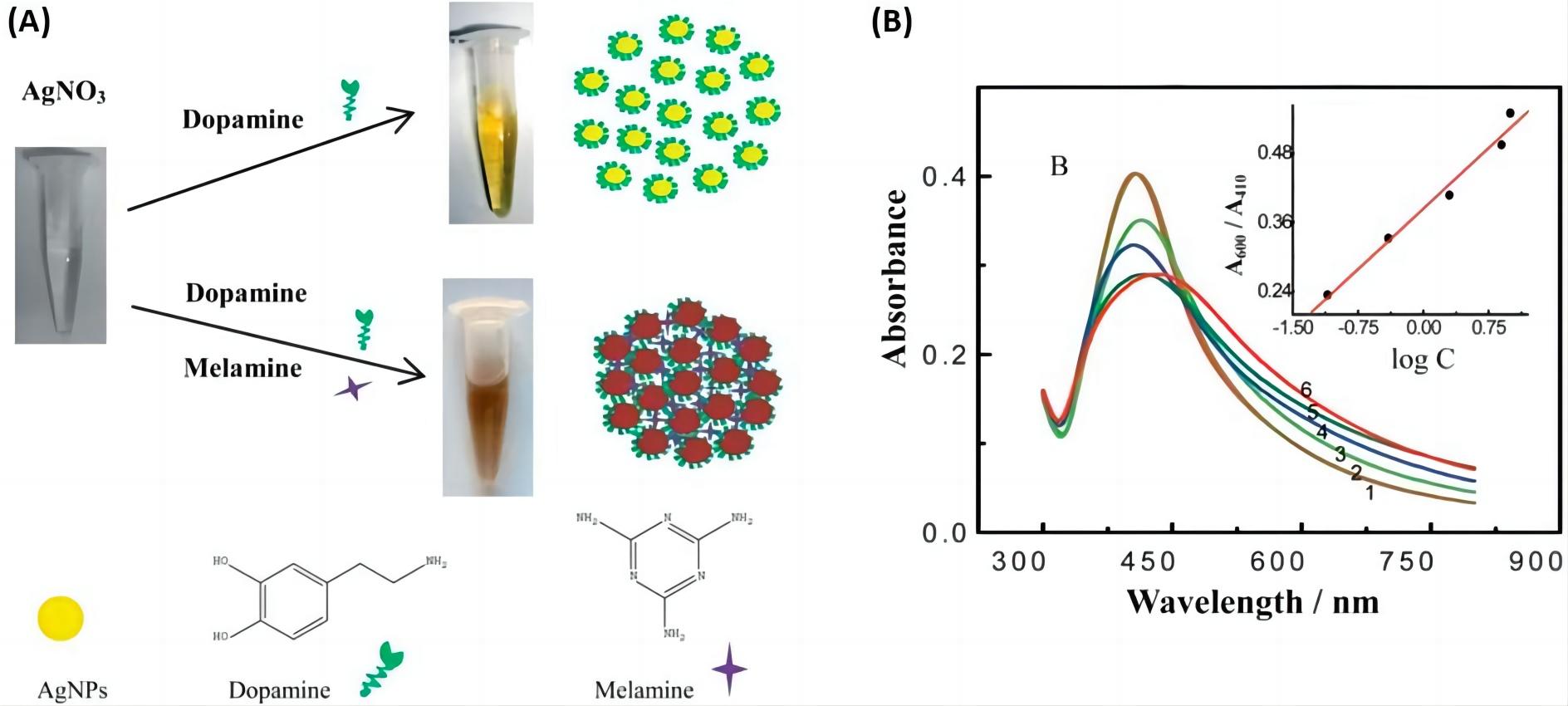 Fig.1 Nanoparticle aggregation-based colorimetric assay for melamine detection using dopamine-modified AgNPs.1, 3
- Colorimetric detection based on destabilization of nanomaterials structure.
The colorimetric detection of pathogens is based on the change and destabilization of nanomaterial structures. Polydiacetylene (PDA) vesicles are one of the most popularly used colorimetric biosensing structures with unique chromatic properties. The blue-colored PDA vesicles are conjugated with pathogen-specific bioreceptors such as antibodies, aptamers, and/or peptides. Upon binding of corresponding pathogens to the bioreceptor-PDA vesicle, a color change from blue to red occurs and can be detected.
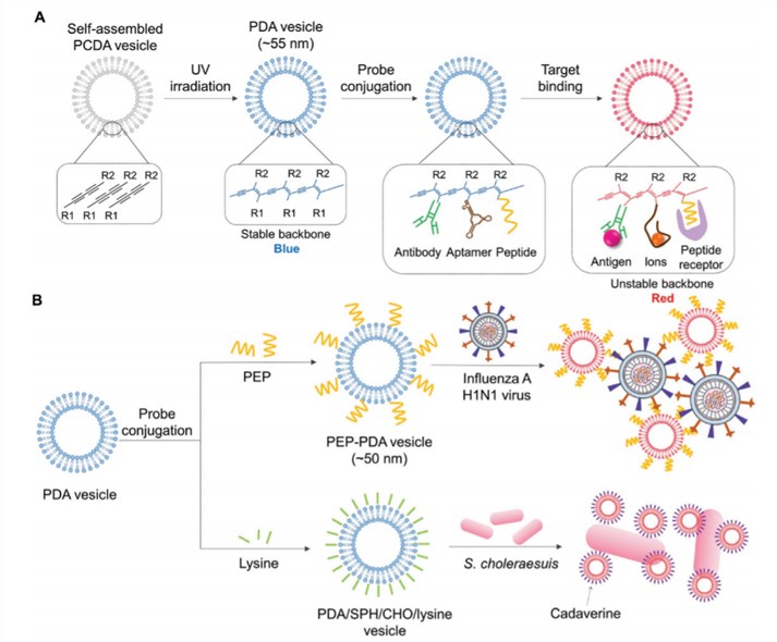 Fig.2 Schematic illustration of colorimetric detection based on destabilization of nanomaterial structure. A) Scheme of a color transition of PDA vesicles by their structure change on target binding. B) Scheme of detecting influenza H1N1 virus with PEP-PDA vesicle system, and detecting S. choleraesuis with PDA/SPH/CHO/lysine vesicle system.2, 3
Our Capabilities
As an expert in the field of IVD development, Creative Biolabs is specialized in flexibly leveraging a variety of biological technologies to support the development of IVD assays to assist. We are committed to offering a full range of IVD assay development services to facilitate disease diagnosis. If you are interested in our services, please contact us to discuss your project and get more details.
References
- Loiseau, Alexis, et al. "Silver-based plasmonic nanoparticles for and their use in biosensing." Biosensors 9.2 (2019): 78.
- Celik, Cagla, et al. "Recent Advances in Colorimetric Tests for the Detection of Infectious Diseases and Antimicrobial Resistance." Diagnostics 13.14 (2023): 2427.
- Distributed under Open Access license CC BY 4.0, without modification.
For Research Use Only.