Custom antibody arrays are an ideal tool for screening a large number of potential biomarkers in a single experiment. This high-throughput capability allows for the rapid identification and validation of diagnostic or prognostic protein markers in complex biological fluids such as serum, plasma, and cerebrospinal fluid, accelerating the path to clinical translation.
Antibody arrays are powerful multiplex immunoassays that allow for the simultaneous detection of hundreds of proteins from a single sample. Creative Biolabs provides comprehensive custom antibody array development services, leveraging our deep expertise to deliver a tailored and highly efficient solution for in vitro diagnostics (IVD) research.
Antibody Array
Antibody arrays, also known as protein microarrays, are an advanced technology for high-throughput protein analysis. They operate on a principle similar to a sandwich ELISA, but on a miniaturized, solid substrate such as a glass slide or a nitrocellulose membrane. In this format, multiple highly specific capture antibodies, each targeting a different protein, are immobilized in discrete spots. This allows for the simultaneous analysis of complex protein mixtures, providing a comprehensive "proteomic snapshot" of a biological sample, which would otherwise require numerous, time-consuming individual assays.
Advantages of Antibody Array
- Exceptional Efficiency: Antibody arrays significantly reduce the time, effort, and cost associated with measuring multiple proteins by eliminating the need for numerous individual Western blots or ELISAs. This consolidated approach streamlines the experimental workflow.
- Minimal Sample Consumption: By analyzing a large number of analytes from a small volume of sample, this technology conserves precious biological material, which is critical for studies with limited tissue or clinical samples.
- High Sensitivity and Broad Dynamic Range: Advanced detection systems and signal amplification techniques, such as Rolling-circle amplification (RCA), enable the detection of both highly abundant and low-concentration proteins, providing a wider quantitative range than traditional methods.
- Enhanced Reliability and Reproducibility: The use of duplicate or quadruplicate spots for each antibody and the incorporation of internal controls ensure a high degree of data redundancy, accuracy, and consistent performance across different experiments and batches.
Our Antibody Array Development Services
Creative Biolabs specializes in designing and manufacturing custom antibody arrays that are perfectly aligned with your unique research objectives. Unlike generic commercial arrays that may not cover all your targets of interest, our service offers complete customization of the array's content. We work closely with you from the initial consultation to meticulously select and validate the optimal antibody pairs, ensuring they are specific and have a high affinity for your target proteins. Our in-house manufacturing process and rigorous quality control protocols guarantee a reliable, high-performance assay that is optimized for your specific sample matrix.
Service Workflow of Antibody Array Development
Our custom antibody array development process is a structured workflow designed to ensure precision and success at every stage.
01Initial Consultation & Project Design
The process begins with a detailed discussion to understand your research objectives, target proteins, and sample types. We collaborate with you to define the optimal project scope, including the selection of antibodies and the final array format.
02Antibody Production & Validation
Based on the project design, our team initiates the development of highly specific, high-affinity capture antibodies. These antibodies undergo a rigorous validation process to confirm their performance and specificity, minimizing the risk of cross-reactivity in the final multiplex assay.
03Array Fabrication
The validated capture antibodies are precisely spotted onto the chosen solid substrate, such as a glass slide or a nitrocellulose membrane, using a microarraying robot. Each antibody is spotted in duplicate or quadruplicate to ensure data redundancy and robust statistical analysis.
04Assay Optimization & Validation
We perform extensive testing to optimize the entire assay protocol for your specific sample matrix. This includes fine-tuning blocking, sample incubation, and detection conditions to maximize signal-to-noise ratio and ensure the array performs robustly and reliably.
05Data Acquisition & Reporting
Once the array is validated, we acquire the data using a high-resolution scanner and perform a comprehensive analysis. We deliver a secure and detailed report that includes all raw data, quality control metrics, and a thorough analysis of the protein expression profiles, providing you with actionable insights for your research.
Applications
Biomarker Discovery and Validation
Pathway and Signaling Profiling
By simultaneously measuring the expression levels and post-translational modifications of multiple key proteins within a signaling cascade, researchers can gain a holistic understanding of cellular responses. This enables the detailed profiling of pathways like apoptosis, inflammation, and cell cycle regulation, revealing novel protein-protein interactions and network dynamics.
Post-Translational Modification Analysis
A key advantage of custom arrays is their ability to specifically profile post-translational modifications (PTMs). By utilizing specific antibodies, we can develop arrays to analyze critical PTMs like phosphorylation and glycosylation, which are essential for understanding protein function and cellular regulation in health and disease.
Immunology and Inflammation Research
Custom arrays are widely used in immunology to profile the expression of a wide range of cytokines, chemokines, and growth factors simultaneously. This provides a comprehensive view of the immune and inflammatory responses, enabling researchers to monitor disease progression, evaluate the efficacy of immunotherapies, and study cellular communication.
Examples of Antibody Array Utility
Rolling-circle amplification (RCA) can greatly enhance fluorescence signals over non-amplified detection, enabling the detection of low-abundance proteins like cytokines.
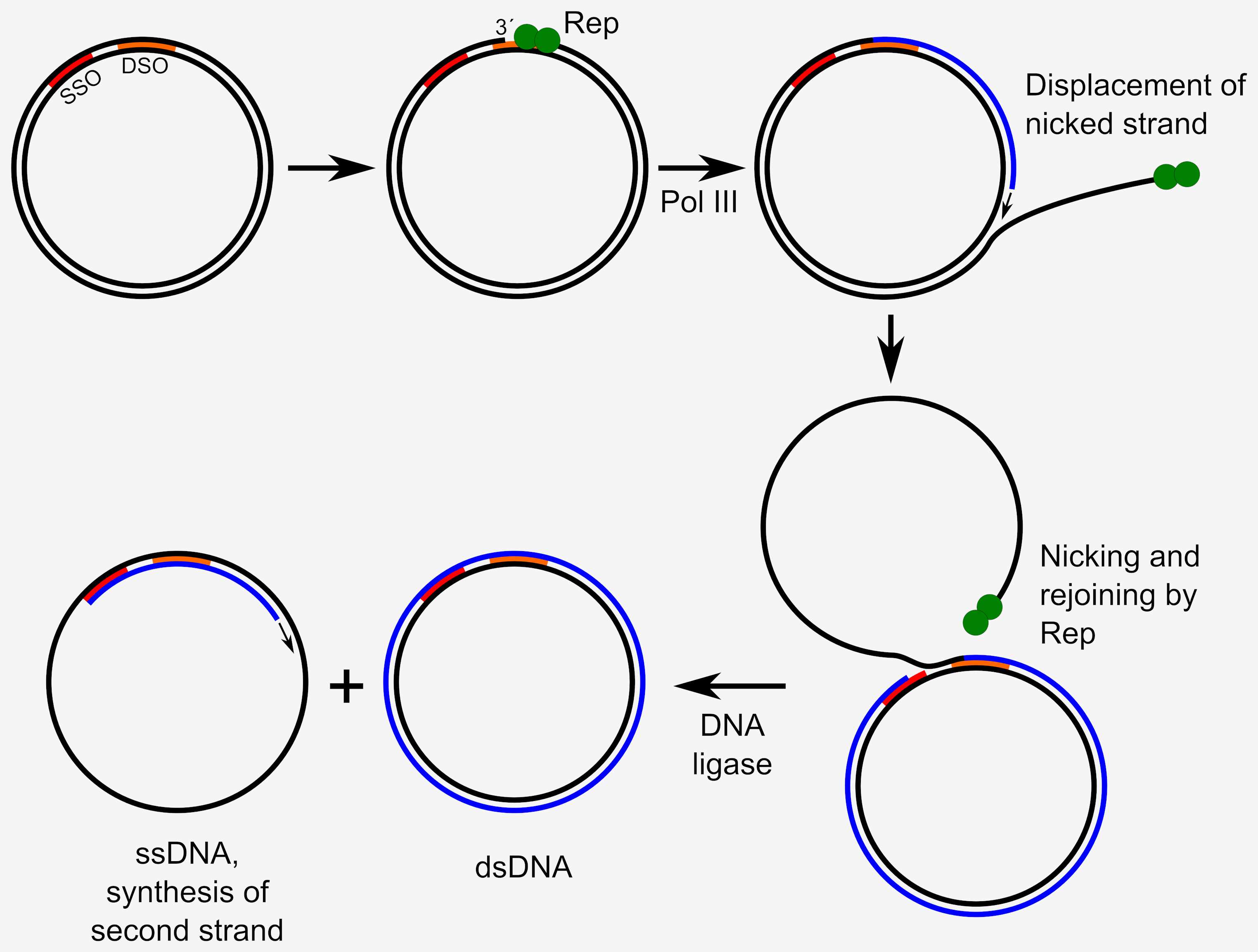 Fig.1 Illustration of RCA. Distributed under CC BY-SA 4.0, from Wiki, without modification.
The biological roles of proteins are determined not just by their abundance, but also by modifications and interactions that occur after translation such as phosphorylation, glycosylation, cleavage, oxidation or reduction, and lipid attachment.
Published Data
1. Development of an Internally Controlled Antibody Microarray
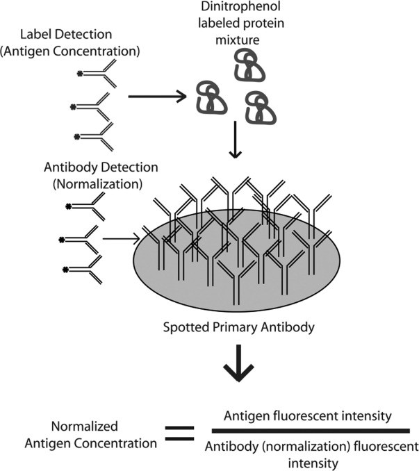 Fig.2 Diagram of the internally controlled antibody microarray.1
In this study, researchers developed a novel antibody microarray for protein expression profiling that used a single antibody and controlled the amount spotted. The array incorporates an internally controlled system where one color represents antibody spot intensity and another indicates antigen levels, allowing for accurate quantification of protein expression. By normalizing the spot intensity, the method reduced variability and lowered detection limits compared to relying on median fluorescence intensity alone. The array was tested with the expression of standard cytokine proteins and cell lysates from mouse macrophages stimulated in vitro, including IL-1β, IL-5, IL-6, and MIP-1α and MIP-1β. Protein expression levels with the antibody microarray were compared with western blot results, showing similar expression patterns, but the antibody array demonstrated superior sensitivity. This technology offers a scalable solution for high-throughput screening of hundreds of proteins in complex biofluids like blood.
2. Developing Multiplexed Antibody Microarray for Quantitative Proteomic
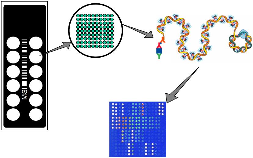 Fig.3 Schematic representation of antibody microarray slide and RCA immunoassay.2
The report detailed the development of first-generation standards for antibody arrays in quantitative proteomics, covering specifications for manufacturing, qualification, assay automation, performance, validation, and data processing. Six dual-antibody sandwich immunoassay arrays were created to measure 170 serum or plasma proteins, experimental procedures were refined through over thirty proteomics studies. Key topics included validating antibody specificity under multiplexed conditions, applying standardized statistical approaches for data analysis, and designing controls for sample normalization. It emphasized stringent quality control, real-time monitoring of assay performance (sensitivity, dynamic range, precision), and procedures to assess the significance of biomarker findings in large-scale studies.
Service Highlights
- Rigorous Quality Control: Our proprietary quality control ensures every capture antibody is meticulously validated for specificity and performance. This minimizes cross-reactivity and background noise, which is essential for precise and reliable data.
- Optimized Protocols: We custom-tailor every aspect of the assay protocol, from sample preparation to detection, to fit your specific research needs. This ensures the array performs optimally with your sample matrix.
- End-to-End Scientific Partnership: We act as your dedicated scientific partner throughout the project. Our PhD-level experts provide consultation on design and offer comprehensive support for data analysis and interpretation, guaranteeing a seamless, successful outcome.
- Flexible Array Formats & Detection: We offer the flexibility of choosing between different array substrates, such as glass slides or membranes. This versatility extends to detection methods, allowing for either fluorescence or chemiluminescence-based assays to suit your experimental requirements.
FAQs
-
Q: How do you ensure the specificity of the antibodies used in your custom arrays?
A: We ensure the specificity through a multi-stage validation process. This involves extensive screening of antibody candidates to confirm their affinity and specificity for the target protein, using techniques like Western blotting and immunofluorescence. We further validate pairs under multiplexed conditions to minimize cross-reactivity, which is crucial for accurate results.
-
Q: What types of samples can be used with the custom antibody array service?
A: A wide variety of biological samples can be used with our custom antibody array service. The service is compatible with common types, including cell lysates, tissue homogenates, serum, plasma, and conditioned media. We meticulously optimize our assay protocols to ensure compatibility and robust performance, which is essential for high-quality and reliable data.
-
Q: Is it possible to include both human and mouse proteins on the same array?
A: Yes, it is possible to include both human and mouse proteins on the same array. Our services are highly flexible and can be designed to include a mix of antibodies specific to different species. This allows us to develop a unique panel to detect a variety of species-specific targets simultaneously, which is useful for comparative studies and xenograft models.
-
Q: How do you handle low-abundance proteins that are difficult to detect?
A: We handle low-abundance proteins by optimizing the assay to maximize sensitivity. This often involves signal amplification techniques, such as RCA or highly sensitive substrates, boosting the signal from low-concentration analytes. Our experts can also adjust assay conditions to ensure the best possible detection limits.
-
Q: Can you develop an array to detect post-translational modifications like phosphorylation?
A: Yes, we can develop an array to detect post-translational modifications. This involves using highly specific antibodies engineered to recognize a particular phosphorylation site. This allows for the precise profiling of these critical modifications, which is essential for understanding cell signaling and disease mechanisms. Our team has extensive experience developing and validating these specialized arrays.
-
Q: Can you help with the experimental design for my study using the array?
A: We are more than capable of helping with experimental design. As part of our end-to-end scientific partnership, we offer consultation on project design to ensure the array is used most effectively. We assist with sample preparation, experimental controls, and data analysis strategies, guaranteeing your study is scientifically sound and poised for success.
If you are interested in our custom antibody array development services, please contact us for more information.
References
- Olle, Eric W., et al. "Development of an internally controlled antibody microarray." Molecular & Cellular Proteomics 4.11 (2005): 1664-1672. Distributed under Open Access license CC BY 4.0, without modification.
- Perlee, L. T., et al. "Development and standardization of multiplexed antibody microarrays for use in quantitative proteomics." Proteome science 2 (2004): 1-22. Distributed under Open Access license CC BY 2.0, without modification.
For Research Use Only.