The pursuit of precise and physiologically representative in vitro models has been a constant objective in cancer research. 2D cell cultures serve as practical tools but fall short of replicating the intricate architecture and tumor microenvironment found in human solid tumors. The 3D cancer spheroid model stands as a revolutionary approach which has proven to be a crucial instrument in cancer biology studies as well as immuno-oncology drug discovery and development, immune response analysis, biomarker discovery and multi-omics analysis. The 3D cancer spheroid designates an aggregate of cancer cells which self-assemble into a spherical formation during culture provided appropriate conditions exist. The cancer spheroids imitate multiple in vivo tumor characteristics including cellular interactions and extracellular matrix dynamics alongside heterogeneous cellular compositions and concentration gradients of oxygen and other vital substances.
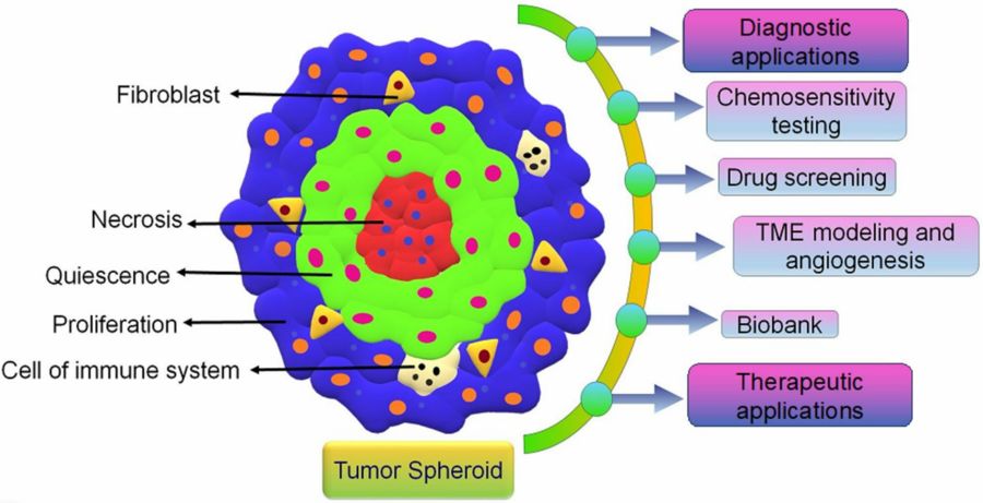 Figure 1 Tumor Spheroid Applications.1,3
What is Cancer Stem Cell Spheroid?
Cancer stem cells (CSCs) are a subpopulation of cells within a tumor that have the ability to self-renew and give rise to all the different cell types within the tumor. Cancer stem cell spheroids represent three-dimensional aggregates which contain a high concentration of CSCs. Scientists create these spheroids by growing tumor cells in media formulations that support CSC growth and self-renewal while typically avoiding adherent surfaces. Cancer stem cell spheroids play a critical role in the processes of tumor initiation and progression as well as metastasis and recurrence. Research in colorectal cancer discovered that cancer stem cell spheroids displayed enhanced tumor initiation capabilities when transplanted into immunodeficient mice than conventional non-stem cell tumor cell populations. Cancer stem cell spheroids demonstrate increased resilience against standard chemotherapy and radiotherapy which provides an explanation for tumor recurrence following treatment. Research into cancer stem cell spheroids enables scientists to discovery novel drug, biomarker, disease research (liver disease, lung disease, breast disease, et al.) and develop targeted treatments which attack these cells and enhance patient survival rates.
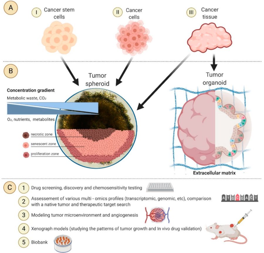 Figure 2 The application of 3D tumor models in personalized medicine.2,3
Spheroid Culture in Cancer Research
Spheroids are essential for cancer research because they accurately replicate in vivo tumor biology. These spheroids demonstrate cellular diversity and gradients that accurately replicate physiological conditions through oxygen and nutrient distribution along with cell-cell communication and extracellular matrix presence which extensively affect both tumor behavior and drug response.
Key advantages of spheroid culture in cancer research include:
-
Mimicking Tumor Microenvironment (TME): The formation of microenvironmental gradients (oxygen, nutrients, pH) and complex interactions between cells and cell-extracellular matrix (ECM) within spheroids establishes essential conditions for tumor development, persistence and resistance to treatment. Traditional cultures provide cells with equal media distribution while failing to establish meaningful cell-cell connections unlike spheroid cultures.
-
Replicating Drug Resistance: In vivo tumors develop resistance because of limited drug penetration to their core regions and changes in cell signaling inside the TME along with quiescent cell populations. Spheroids show restricted drug penetration into their core areas and develop multi-drug resistance patterns similar to those seen in living tumors. Research indicates that drug effectiveness in spheroids demonstrates stronger alignment with in vivo xenograft models than with traditional 2D cell cultures.
-
Studying Cell Proliferation and Viability: Spheroids contain unique proliferative areas which enable detailed research of anti-proliferative drugs because they allow researchers to differentiate between rapidly dividing cells on the surface and dormant cells inside.
-
Investigating Metastasis and Invasion: Researchers employ spheroid models to investigate cellular behaviors like migration and invasion in surrounding matrices which helps to understand the mechanics of metastasis.
-
High-Throughput Screening (HTS) Compatibility: Initially considered complicated for high-throughput applications, new spheroid culture methods and automated imaging technology now allow 3D spheroids to be used effectively for rapid drug screening across extensive compound libraries.
Studying 3D cancer spheroids requires visualization to comprehend their structure and function. Researchers have created advanced imaging methods to analyze 3D cancer spheroids at various scales. Confocal microscopy stands as one of the standard methods that enables researchers to capture high-resolution images of spheroid internal structures. Researchers can use this method to obtain precise details about interactions between cells within the spheroid as well as the spatial distribution of various cell types and targeted protein locations. Researchers used confocal microscopy to study the distribution of epithelial-mesenchymal transition proteins within 3D breast cancer spheroids which revealed differential protein expression between outer and inner spheroid layers. Imaging of 3D cancer spheroids involves various techniques:
|
Imaging Technique
|
Description
|
Primary Applications
|
|
Brightfield Microscopy
|
Standard microscopic visualization using transmitted light.
|
Basic observation of spheroid formation, morphology, and growth over time.
|
|
Fluorescence Microscopy
|
Uses fluorescent probes/dyes that emit light upon excitation.
|
Assessing cell viability (live/dead stains), proliferation rates (EdU), apoptosis markers, and general protein expression within spheroids.
|
|
Confocal Microscopy
|
Provides high-resolution optical sections in 3D by rejecting out-of-focus light.
|
Detailed visualization of cellular organization, protein localization, and drug penetration in 3D without physical sectioning.
|
|
High-Content Imaging (HCI)
|
Automated systems for rapid acquisition and analysis of images from multi-well plates.
|
Quantitative assessment of spheroid growth, compactness, viability, apoptosis, and specific biomarker expression in a high-throughput manner for drug screening.
|
Drug Combination in Cancer Spheroids
Research into cancer treatment shows great potential when drug combinations are applied to cancer spheroids. Tumors develop resistance to single-drug therapies because cancer represents a complex disease. Researchers can determine more effective drug combinations against cancer cells by testing drugs in 3D cancer spheroid models because these combinations demonstrate superior performance compared to individual drugs. A research study on ovarian cancer examined 3D spheroids of ovarian cancer cells that received treatment from both chemotherapy and targeted therapy drugs. The combination therapy demonstrated a significantly stronger suppression of spheroid growth and triggered more cell death than treatments with individual drugs. The combination drugs attack various tumor-related pathways resulting in cancer growth inhibition and resistance prevention which single-drug treatments cannot achieve. 3D cancer spheroid models are crucial tools in drug discovery, particularly in areas like liver cancer, lung cancer, breast cancer and immuno-oncology. They are also invaluable for analyzing immune response, assessing drug toxicity and conducting multi-omics analyses.
Why Spheroids are Superior for Combination Therapy Evaluation?
-
Mimicking Drug Penetration Barriers
Drug penetration in solid tumors when studied in vivo shows that well-perfused outer layers receive adequate drug delivery while the hypoxic inner core obtains much lower concentrations. The open access to drugs that characterizes 2D monolayers prevents them from replicating the drug penetration limitations observed in solid tumors. The penetration barriers of drugs in larger spheroids (>200-300 µm) replicate those found in actual tumor environments. The majority of drugs stay within the outer layers of cells which reflects the same pattern seen in patient tumors. Achieving therapeutic effectiveness across a spheroid calls for greater drug concentrations and ideally utilizes drug combinations.
-
Heterogeneous Cell Populations
Spheroid zones that include proliferative, quiescent and hypoxic cells lead to varying responses to drugs. One drug may show high effectiveness against outer layer proliferating cells yet fail to impact the quiescent or hypoxic cells located in the core. Designing drug combinations enables simultaneous targeting of multiple cell populations which results in more effective elimination of cells.
-
Overcoming Resistance Mechanisms
The 3D tumor architecture and microenvironment play a significant role in determining drug resistance mechanisms in vivo. Spheroids provide superior modeling for complex resistance pathways such as altered drug metabolism and increased efflux pump activity which are influenced by cell-cell and cell-ECM interactions than 2D cultures. Testing drug combinations designed to bypass or reverse resistance mechanisms achieves greater effectiveness when conducted on spheroids.
Frequently Asked Questions (FAQs)
Q: What is the primary advantage of 3D cancer spheroids over 2D cell cultures?
A: 3D spheroids better replicate the in vivo tumor microenvironment because they include accurate cell-cell interactions along with extracellular matrix components and oxygen and nutrient gradients. Drug responses from 3D spheroids produce more accurate physiological outcomes and better predict in vivo efficacy than 2D monolayers.
Q: How do 3D spheroids demonstrate drug resistance differently from 2D cultures?
A: Spheroids evolve internal gradients that obstruct drug entry into their center and mimic the characteristics of solid tumors in a living organism. Within spheroids exist dormant and oxygen-deprived cells that conventional treatments cannot easily affect which results in stronger drug resistance than in uniformly treated 2D culture cells.
Q: It's possible to apply high-throughput screening (HTS) to 3D cancer spheroids?
A: The development of ultra-low attachment plates in formats like 96- or 384-wells together with automated high-content imaging systems has enabled 3D cancer spheroids to become highly suitable for HTS applications which facilitates effective screening of large compound libraries.
What Creative Biolabs Can Provide?
Creative Biolabs delivers complete biomedical research services by specializing in 3D cancer spheroid modeling which supports drug discovery and both toxicity and fundamental cancer biology investigations. Our deep expertise and advanced platforms enable us to deliver customized solutions which represent in vivo tumor complexity to help our clients speed up their research and development processes.
Custom Services
-
Custom 3D Spheroid Generation: Produces reproducible 3D spheroids from diverse cell types, including cancer lines and patient-derived cells, customized for specific research needs.
-
Co-culture and Complex Spheroid Models: Create and analyze co-culture spheroids with varied cell types to accurately simulate specific tissue microenvironments.
-
3D Spheroid Based Biomarker Discovery: Services for generating and analyzing PDTS, offering a robust platform for drug response evaluation.
-
3D Spheroid Based Drug Discovery: Tumor spheroids are favored for drug screening as they effectively replicate in vivo drug penetration barriers and resistance mechanisms.
-
3D Spheroid Based Toxicity Evaluation: Spheroids serve as a more reliable platform for evaluating drug-induced toxicity, yielding better predictions of in vivo adverse effects than 2D models.
Culture Products
3D Spheroid Model
Creative Biolabs provides comprehensive biomedical research services, focusing on 3D cancer spheroid modeling to aid in drug discovery, toxicity testing, and fundamental cancer biology studies. Contact us today to learn more!
References
-
Nayak P, Bentivoglio V, Varani M, et al. Three-dimensional in vitro tumor spheroid models for evaluation of anticancer therapy: recent updates. Cancers, 2023, 15(19): 4846. https://doi.org/10.3390/cancers15194846
-
Yau J N N, Adriani G. Three-dimensional heterotypic colorectal cancer spheroid models for evaluation of drug response. Frontiers in Oncology, 2023, 13: 1148930. https://doi.org/10.3389/fonc.2023.1148930
-
Distributed under Open Access license CC BY 4.0, without modification.
Research Model
Related Sections: