Lots of the properties of the aptamer make them better imaging ligands than antibodies for effective clinical cancer diagnosis and tracking. Aptamers conjugated with radioisotopes have been actively exploited in computed tomography (CT), positron emission tomography (PET), and single-photon emission computed tomography (SPECT) for detecting both primary tumor and metastases. Having accumulated rich experience in imaging research, Creative Biolabs provides customized aptamer-based PET/SPECT imaging tool development services. Besides, of-the-sheIf products including antibody or aptamer-based products are available for you.
Imaging with Aptamer
Nuclear imaging (including PET and SPECT) can monitor the biological events deep in the body and provide a longitudinal assessment of the same patient with high detection sensitivity. Combined with the synthesis of radioactive molecules with increased specificity for different biological targets, the development of PET and SPECT imaging modalities promoted the field of nuclear medicine into a new era. Aptamers selected from SELEX have shown high sensitivity and specificity required for nuclear imaging probes.
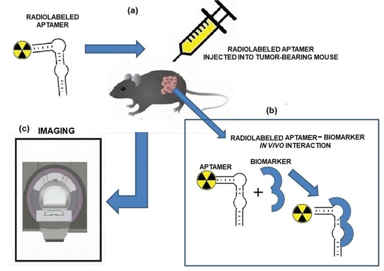 Fig.1 Schematic representation of the pre-clinical use of radiolabeled aptamers. (Filippi, 2020)
PET Imaging
When compared with SPECT, PET can provide higher sensitivity. PET takes full advantage of the unique γ-ray emissions from the decay of a positron. The ring detector of PET can monitor the annihilation events and convert the signals into sonograms, which will eventually be reconstructed to produce tomographic images. Due to its decay characteristics and easy attachment to ligands with high affinity, 64Cu, 11C, 18F, 68Ga represent attractive radiotracers that have broadly applied for in vivo PET imaging. Aptamer-modified nanomaterials are readily applicable for PET imaging.
SPECT Imaging
SPECT imaging relies on the detection of γ-ray emissions from the radioisotopes by a gamma camera. The most common isotopes for SPECT imaging include 99mTc, 111In, 123I, and 131I. One earlier successful aptamer-based imaging system was to use a radiolabeled 99mTc aptamer NX21909 specifically targeting human neutrophil elastase in identifying inflammation sites in a rat model of reverse passive Arthus reaction.
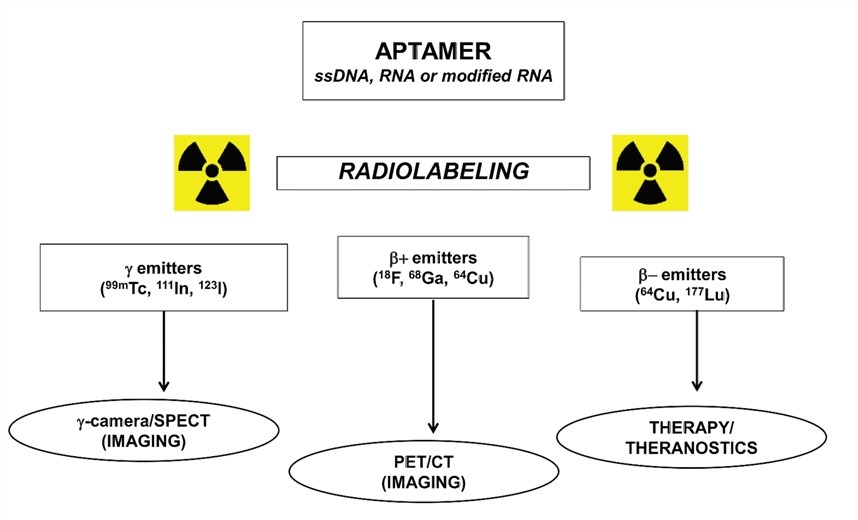 Fig.2 Main radionuclides utilized for aptamer-radiolabeling. (Filippi, 2020)
Services at Creative Biolabs
Aptamer-based molecular imaging has been applied to image a broad range of target molecules. Most of the imaging studies are done in the context of cell culture and animal models.
Applicable Scope
- Sense extracellular signaling molecules
- Protein localization and trafficking in cells
- RNA imaging
- Small molecule imaging
Services
- Radiotracer-labeled aptamer development
- Tailored labeling methods
- Aptamer functionalization
- Aptamer conjugation optimization
- Biodistribution analysis
If you are interested in aptamer-based imaging tool development, please feel free to contact us for more information.
Reference
- Filippi, L.; et al. Aptamer-based technology for radionuclide targeted imaging and therapy: a promising weapon against cancer. Expert Review of Medical Devices. 2020, 17(8): 751-758.
For Research Use Only.