Lens-free (or lens-less) imaging is emerging as a cost-effective, compact, and lightweight detection method that can serve numerous biological applications. In lens-less microscopy, a diffraction pattern resulting from an object is recorded directly on a digital image sensor array without being optically imaged or magnified by any lens elements. Lens-free imaging can generate high-resolution images within a field-portable platform, which is ideal for affordable point-of-care devices aiming at resource-limited settings.
The simplest form of lens-less imaging is shadow imaging. In shadow imaging, a transparent sample is illuminated with a spatially limited light source, and the shadow of the sample is recorded directly on the image sensor.
Advantages of Lens-less Imaging
Compared with conventional lens-based optical microscopes, lens-less imaging has several key advantages.
- Simplicity, lightweight, and compactness: A lens-free imaging system can be easily constructed using a light source, an aperture (pinhole), and an image sensor, making the system compact (a few cubic centimeters) and lightweight and hence portable, which is suitable for point-of-care (POC) diagnostic systems.
- Cost-effectiveness: The main components of a lens-free imaging system include off-the-shelf items such as a commercial light-emitting diode (LED) and a complementary metal-oxide-semiconductor (CMOS) image sensor, making it an affordable imaging technology.
- Three-dimensional (3D) imaging: lens-free imaging can provide a rapid way of screening large sample volumes in 3D, providing a high-throughput microscopy platform.
Lens-Less Shadow Imaging in Cell Classification
The classification of white blood cells (WBCs) is a basic and important method to diagnose diseases in medical fields. The traditional method uses a blood smear and Wright’s staining, and the different types of white cells are counted under the objective lens of a microscope, which is time-consuming.
To make a smaller, more automatic and low-cost blood cell analyzer, much research has focused on lens-less shadow imaging. Scientists proposed a WBC classification method based on shadow imaging of a complementary metal-oxide-semiconductor (CMOS) sensor. For the blood tests from whole blood samples from six outpatients (proportion of WBCs), the result obtained was a mean correlation index of 0.96. The mean error of the percentage of neutrophils was 3.45%, monocytes was 6.04%, and lymphocytes was 6.7%. This method was not only cheaper but also more automated.
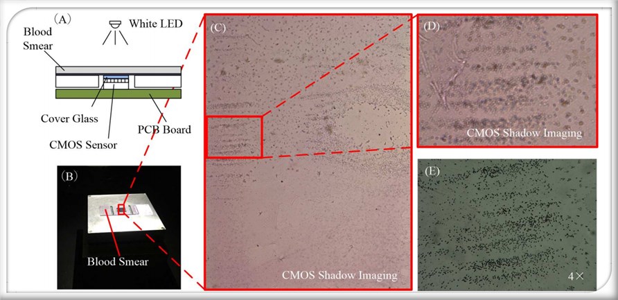 Fig.2 Shadow imaging system based on CMOS technology.1
Lens-less Color Shadow Imaging in Pathology Diagnosis
In a study, to demonstrate the potential application of the color shadow imaging prototype for pathology evaluation, the muscle tissue specimen sample is imaged by scientists. The image of the whole sample can hardly be captured for the traditional microscope with the high-magnification lens, while the resolution-enhanced, lens-less color shadow imaging microscopy (RELCSIM) can easily achieve the field-of-view (FOV). The proposed system promises to greatly improve the efficiency of pathology diagnosis.
Creative Biolabs not only provides best-in-class services but also offers a range of solutions to carefully adapt to each project's specifications. Our professional team is also willing to share long-standing experience with our customers. If you want to know more information, please directly contact us.
Reference
- Fang, Yuan, et al. "An on-chip instrument for white blood cells classification based on a lens-less shadow imaging technique." PloS one 12.3 (2017): e0174580. Distributed under Open Access license CC BY 4.0, without modification.
For Research Use Only.